

Cambridge International AS and A Level Biology 9: Gas exchange
Cambridge International Biology 9700 notes on airways, alveoli, tissue recognition, airway defence and alveolar gas exchange.
Gas Exchange is Cambridge International Biology 9700 Topic 9. The current official boundary covers the lungs, conducting airways, alveoli, capillary networks, tissue distribution and structure-function relationships. It does not list smoking-related disease or detailed ventilation mechanics in this topic. Theory owns recognition and explanation; microscope handling and biological drawing execution remain in the dedicated practical hub.
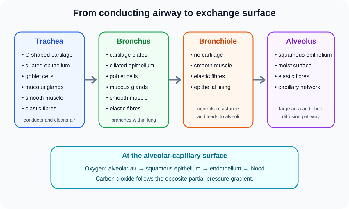
1. The route from air to blood
Air enters the gas exchange system and travels through the trachea, bronchi and progressively smaller bronchioles before reaching alveoli. The lungs contain the branching airways, alveoli and an extensive capillary network.
The trachea is a large conducting airway. It divides into bronchi, which enter the lungs and branch repeatedly. Bronchioles are smaller airways within the lungs and lead toward the alveolar region. Alveoli are microscopic air spaces where oxygen and carbon dioxide are exchanged with capillary blood.
Conducting airways move and condition air. Alveoli provide the exchange surface. A description that calls the trachea itself the main diffusion surface confuses these roles.
2. Trachea
The tracheal wall contains C-shaped cartilage, smooth muscle, elastic fibres, mucous glands and a lining with ciliated epithelial cells and goblet cells. Cartilage keeps the airway open while its incomplete ring permits flexibility and allows the oesophagus behind it to expand during swallowing.
Goblet cells and mucous glands secrete mucus. The mucus traps dust and microorganisms. Cilia beat to move the contaminated mucus toward the throat, reducing the chance that particles and pathogens reach delicate alveolar surfaces.
In transverse section, a plan diagram should show the lumen and relative tissue layers but not individual epithelial cells. Cellular recognition questions can ask for ciliated cells or goblet cells at higher magnification.
3. Bronchi
Bronchi share several tissues with the trachea: cartilage, smooth muscle, elastic fibres, ciliated epithelium, goblet cells and mucous glands. Their cartilage is commonly arranged as irregular plates rather than one large C-shaped ring.
Cartilage maintains patency, while smooth muscle changes airway diameter and elastic fibres support recoil. Mucus and ciliary action continue the airway-cleaning mechanism. As bronchi branch and become smaller, the relative appearance and distribution of tissues change.
A bronchus can be distinguished from a blood vessel because it has an epithelial airway lining and often cartilage plates. A thick wall alone is not enough for identification.
4. Bronchioles
Bronchioles have small lumens and no cartilage. Their walls contain smooth muscle and elastic fibres. Ciliated epithelium may be present, especially in larger bronchioles, while mucus-producing structures become less prominent along the branching pathway.
Check this topic from memory
Attempt the matching topic bank before reopening the notes. Use each missed idea to decide what to review next.
Start the topic quiz