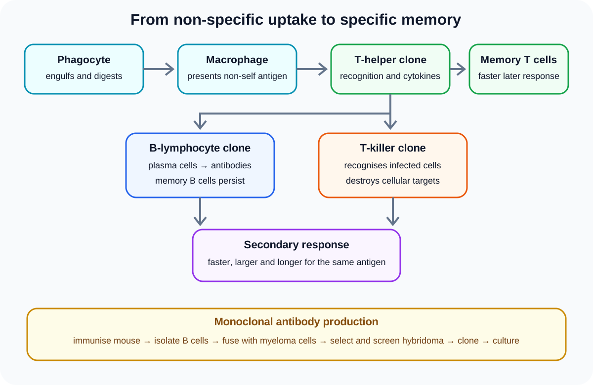
Immunity is Cambridge International Biology 9700 Topic 11. It links phagocytes and antigen recognition to primary and secondary immune responses, antibody structure, monoclonal antibodies, active and passive immunity, vaccines and population-level disease control. Theory owns immune mechanisms and interpretation; assay execution and experimental evaluation remain in the dedicated practical hub.
1. Phagocytes
Neutrophils and macrophages are phagocytes. They respond to chemical signals, recognise foreign material and surround a pathogen with their cell-surface membrane. The pathogen becomes enclosed in a phagosome.
Lysosomes fuse with the phagosome and release hydrolytic enzymes. These digest the pathogen. Useful products may enter the cytoplasm, while residues can be removed by exocytosis. Phagocytosis is non-specific because the same general process acts against many different foreign targets.
Neutrophils are short-lived cells that can reach infected tissue rapidly. Macrophages are larger, can remain in tissues and can present pathogen antigens after digestion. Both engulf material, but their roles are not identical.
2. Self and non-self antigens
An antigen is a molecule, often a protein or glycoprotein, that is recognised as foreign and can stimulate a specific immune response. Antigens have particular shapes that interact with complementary receptors or antibody binding sites.
Self antigens are normal molecules on a person's own cells and are usually tolerated by the immune system. Non-self antigens are recognised as foreign. They may occur on a pathogen, infected cell, transplanted tissue or other foreign material.
An antigen is not the whole bacterium or virus. It is a molecular feature that can be recognised. One pathogen may carry several different antigens and therefore stimulate several lymphocyte clones.
3. Antigen presentation and T-helper activation
After a macrophage digests a pathogen, it displays fragments of non-self antigen on its surface. A T-helper lymphocyte with a complementary receptor binds to the presented antigen. This selects the matching cell from many lymphocyte specificities.
The selected T-helper cell divides by mitosis, producing a clone. Activated T-helper cells release cytokines that stimulate matching B-lymphocytes and T-killer lymphocytes. Specificity depends on complementary recognition, not on the immune system designing a new receptor after infection.
4. B-lymphocytes and antibodies
A B-lymphocyte with a complementary surface receptor binds the antigen and, with appropriate T-helper signalling, undergoes clonal expansion. Some cells differentiate into plasma cells. Plasma cells have extensive rough endoplasmic reticulum and secrete large quantities of one antibody specificity.
Check this topic from memory
Attempt the matching topic bank before reopening the notes. Use each missed idea to decide what to review next.
Antibodies bind their complementary antigens. Binding can neutralise a toxin or pathogen attachment site, aggregate antigens or mark foreign material for phagocytosis. The antibody does not usually digest the pathogen itself.
Other cells in the clone become memory B cells. They remain after the primary response and permit a faster response if the same antigen is encountered again.
5. T-killer cells
T-killer lymphocytes recognise specific non-self antigen displayed by infected body cells. After activation and clonal expansion, they destroy the infected cells, limiting sites where intracellular pathogens can reproduce.
T-killer cells act on infected host cells rather than producing antibodies. Their action complements the antibody response, which acts on accessible antigens outside cells or on cell surfaces.
Some activated T cells become memory cells. The syllabus limits named T-cell types here to T-helper and T-killer cells, so additional catalogues are unnecessary.
6. Primary, secondary and long-term responses
The primary immune response follows first exposure to an antigen. Selecting and expanding the rare complementary lymphocyte clones takes time, so antibody concentration rises after a delay and may reach a moderate peak before declining.
Memory B and T cells remain. On later exposure to the same antigen, they divide and differentiate rapidly. The secondary response begins sooner, reaches a higher antibody concentration and commonly persists longer. The pathogen may be controlled before noticeable disease develops.
Memory is antigen-specific. Immunity to one antigen does not guarantee protection against an unrelated pathogen or a substantially changed antigen.
7. Antibody structure and function
An antibody is a globular protein made of two identical heavy polypeptide chains and two identical light chains joined by disulfide bonds. The four chains form a Y-shaped molecule. Variable regions at the ends form two antigen-binding sites with shapes complementary to a particular antigen.
Two binding sites allow one antibody to connect antigens and contribute to agglutination. A flexible hinge lets the arms bind sites at different angles. Constant regions are shared within an antibody class and interact with other components of the immune system, including phagocytes.
Specificity comes from variable-region amino-acid sequence and three-dimensional shape. Disulfide bonds stabilise the molecule; they do not determine antigen specificity by themselves.
8. Hybridomas and monoclonal antibodies
To produce monoclonal antibodies, a mouse is exposed to the target antigen so matching B-lymphocytes form. Antibody-producing B cells are collected, commonly from the spleen, and fused with myeloma cells. The B cell contributes the antibody specificity; the tumour cell contributes the ability to divide repeatedly.
The resulting hybridomas are selected and screened to find a cell producing the required antibody. That hybridoma is cloned, cultured and used to produce a large supply of identical antibodies. Because the cells descend from one selected hybridoma, the antibodies bind the same epitope.
Screening matters: fusion alone creates many cell lines, not all of which produce the required antibody.
9. Monoclonal antibodies in diagnosis and treatment
In diagnosis, a monoclonal antibody binds a specific target antigen. A linked marker, such as an enzyme or fluorescent molecule, makes binding detectable. The presence or amount of signal can reveal a pathogen molecule, hormone or disease-associated marker. Controls are needed because a signal depends on assay design as well as antibody specificity.
In treatment, a monoclonal antibody can bind a selected cell-surface receptor or antigen, block a signalling interaction, mark target cells for immune action or deliver a linked drug to cells carrying the target. Specific targeting can reduce effects on non-target cells, but only when the target is sufficiently selective.
10. Active and passive immunity
Active immunity develops when a person's lymphocytes respond to an antigen and produce effector and memory cells. Natural active immunity follows infection; artificial active immunity follows vaccination. It develops relatively slowly but can be long-lasting because memory cells form.
Passive immunity results from receiving antibodies made by another organism. Natural passive immunity includes maternal antibodies transferred across the placenta or in breast milk. Artificial passive immunity follows injection of prepared antibodies. Protection is immediate but temporary because the recipient did not form matching memory cells.
Natural and artificial describe how exposure occurs. Active and passive describe who produced the antibodies and whether the recipient mounted the response.
11. Vaccination and disease control
A vaccine contains pathogen antigens in a form intended to stimulate an immune response without causing the full disease. The antigens select matching lymphocytes, producing plasma cells, antibodies and memory cells. Later exposure can then trigger a rapid secondary response and long-term protection.
Vaccination programmes reduce the number of susceptible hosts and can interrupt chains of transmission. When enough people are immune, an infected person is less likely to encounter a susceptible host. This indirect protection is especially important for people who cannot receive a particular vaccine or do not develop strong immunity.
Population control depends on coverage, vaccine effectiveness, duration of protection, pathogen transmission and antigen change. Vaccination reduces risk; it does not make transmission mathematically impossible in every context.
Worked application: interpreting two antibody curves
A student receives a vaccine on day 0 and a booster on day 42. Antibody concentration rises slowly after the first dose because complementary B and T cells must be selected, cloned and differentiated. It then falls, while memory cells persist. After the booster, concentration rises sooner, reaches a higher peak and remains elevated longer because memory cells respond rapidly and form many plasma cells. If an unrelated antigen is introduced, the existing memory cells do not produce the same accelerated response because their receptors are not complementary. The graph therefore supports antigen-specific memory rather than a permanent non-specific increase in all immunity.
Common misconceptions and corrections
Calling phagocytosis antigen-specific. It is an immediate non-specific response.
Saying lysosomes engulf pathogens. The cell membrane forms the phagosome; lysosomes fuse with it.
Calling an antigen the whole pathogen. It is a recognised molecular feature.
Saying all self antigens trigger attack. They are normally tolerated.
Saying macrophages only digest material. They can also present antigen.
Saying T-helper cells make antibodies. Plasma cells secrete antibodies.
Saying B cells are manufactured after infection. Existing complementary clones are selected.
Calling plasma cells memory cells. They are specialised antibody-secreting effectors.
Saying antibodies digest pathogens. They bind antigens and enable specific effects.
Saying T-killer cells secrete antibodies. They destroy infected host cells.
Calling the primary response faster than the secondary. Clone selection makes it slower.
Saying memory protects against every antigen. Memory is specific.
Giving an antibody one binding site. It has two equivalent antigen-binding sites.
Saying the constant region determines specificity. Variable regions form the binding sites.
Calling a hybridoma a fused pair of two B cells. It combines a B cell and a myeloma cell.
Skipping hybridoma screening. The required specificity must be identified.
Saying monoclonal means many different antibodies. The antibodies share one specificity.
Calling vaccination passive immunity. It stimulates the recipient's active response.
Saying passive immunity forms memory cells. Supplied antibodies give temporary protection.
Claiming vaccination always prevents every transmission event. Programme effects depend on coverage and biology.
Assessment guidance
Write immune-response sequences in causal order: phagocytosis, antigen presentation, complementary recognition, clonal expansion, effector cells and memory. Keep macrophage, T-helper, T-killer, B-cell and plasma-cell roles distinct. For antibody questions, connect each structural feature to a functional consequence rather than drawing an unlabeled Y. Hybridoma answers need immunisation, B-cell isolation, fusion, selection, screening, cloning and culture. Immunity classifications require both axes: active or passive and natural or artificial. Vaccination discussions should move from antigen exposure to memory and then from fewer susceptible hosts to reduced transmission, with realistic limits.
Retrieval practice
Trace one pathogen from engulfment to antigen presentation and parallel B-cell and T-killer responses. Draw and annotate an antibody with chains, bonds, variable regions, binding sites, hinge and constant regions. Reconstruct hybridoma production in order and state the contribution of each fused cell. Classify four immunity examples on a two-axis grid. Finally, sketch primary and secondary antibody curves and explain how a vaccination programme changes transmission at population level.