Homeostasis is Cambridge International Biology 9700 Topic 14. It connects negative feedback to kidney function, osmoregulation, glucagon signalling, blood-glucose control, enzyme-based glucose measurement and guard-cell responses. Theory owns mechanisms, structures and interpretation; biosensor operation, specimen work and investigation execution remain in the dedicated practical hub.
1. Homeostasis and negative feedback
Homeostasis is the maintenance of a relatively stable internal environment within limits despite internal or external change. Stable temperature, blood glucose concentration and blood water potential support enzyme activity, membrane function and cell metabolism.
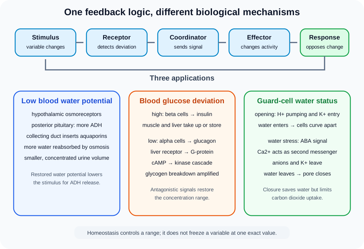
A stimulus is a detected change. Receptors monitor the variable, coordination systems process information, and effectors such as muscles or glands produce a response. In negative feedback, the response opposes the original deviation. The variable fluctuates around a range rather than remaining perfectly fixed.
Both nervous and endocrine systems can coordinate responses. The speed, signal type and target differ, but each can link a detected condition to effectors.
2. Urea and kidney organisation
Excess amino acids cannot be stored as amino acids indefinitely. In the liver, their amino groups are removed by deamination and nitrogen-containing waste is converted to urea. Blood carries urea to the kidneys for excretion.
Each kidney has a tough fibrous capsule, an outer cortex, an inner medulla and a renal pelvis leading to the ureter. Branches of the renal artery bring blood for filtration, while branches of the renal vein return blood after processing.
Nephrons extend through cortex and medulla. The glomerulus and Bowman's capsule lie in the cortex. The proximal convoluted tubule and distal convoluted tubule are cortical. Loops of Henle extend into the medulla, and collecting ducts pass through the medulla toward the renal pelvis.
3. Nephron blood supply and route
An afferent arteriole supplies the glomerular capillaries and an efferent arteriole drains them. The efferent vessel leads to capillaries associated with the tubule. This arrangement supports filtration followed by reabsorption.
Filtrate follows Bowman's capsule, proximal convoluted tubule, loop of Henle, distal convoluted tubule and collecting duct. Blood and filtrate are separate streams. Useful substances can move from tubule fluid into tissue fluid and then nearby capillaries.
Recognition questions may use diagrams, photomicrographs or electron micrographs. Use epithelial form, lumen, location and neighbouring structures rather than one memorised outline.
Check this topic from memory
Attempt the matching topic bank before reopening the notes. Use each missed idea to decide what to review next.
The glomerulus receives blood at high hydrostatic pressure. The afferent arteriole is wider than the efferent arteriole, helping maintain this pressure. Water and small solutes are forced from blood through the filtration barrier into Bowman's capsule.
The barrier includes fenestrated capillary endothelium, a basement membrane and filtration slits between podocyte processes. Blood cells and most large plasma proteins remain in capillaries. Water, ions, glucose, amino acids and urea can enter glomerular filtrate.
Ultrafiltration is pressure-driven and size-selective. It is not active transport and does not selectively retain every useful small molecule. Selective recovery occurs later.
5. Selective reabsorption in the proximal tubule
The proximal convoluted tubule reabsorbs all glucose and amino acids under normal conditions, along with much water and many ions. Sodium-potassium pumps in the basal membranes use ATP to lower sodium concentration inside epithelial cells. Sodium then enters from the tubule lumen through cotransport proteins with glucose or amino acids.
Glucose and amino acids leave toward tissue fluid through membrane proteins and enter capillaries. Water follows solute movement by osmosis. The exact movements depend on membrane surfaces and gradients, so do not describe every substance as actively pumped directly from filtrate to blood.
Proximal-tubule cells have microvilli for large surface area, many mitochondria for ATP supply, infolded basal membranes and a thin epithelial route. These features support high transport capacity.
6. Osmoregulation and ADH
Osmoregulation controls blood water potential. Osmoreceptors in the hypothalamus detect a decrease in water potential. The posterior pituitary gland releases more antidiuretic hormone, ADH, into the blood.
ADH binds to receptors on collecting-duct cells and causes more aquaporin water channels to be inserted into their cell-surface membranes. Collecting ducts become more permeable to water. More water leaves the filtrate by osmosis into the medulla and returns to blood, producing a smaller volume of more concentrated urine.
When blood water potential rises, less ADH is released, fewer aquaporins are present and less water is reabsorbed, producing more dilute urine. Restoring water potential reduces the original stimulus, completing negative feedback.
7. Glucagon as a cell-signalling example
When blood glucose concentration falls, glucagon binds to a complementary cell-surface receptor on a liver cell. Receptor conformation changes and activates a G-protein. The G-protein stimulates adenylyl cyclase, which forms cyclic AMP, cAMP, as a second messenger.
cAMP activates protein kinase A. This initiates a phosphorylation cascade in which each activated enzyme can activate many molecules at the next stage. The signal is amplified. The final activated enzyme promotes breakdown of glycogen, releasing glucose for export from the liver.
Glucagon does not need to enter the cell. An extracellular signal is converted into a large intracellular response through receptor binding, second-messenger formation and enzyme amplification.
8. Blood-glucose negative feedback
When blood glucose rises, pancreatic beta cells release insulin. Insulin increases glucose uptake by muscle cells and promotes glucose use. In liver and muscle, it promotes conversion of glucose to glycogen. Liver cells also reduce glucose output, so blood concentration falls toward its range.
When blood glucose falls, pancreatic alpha cells release glucagon. Glucagon acts mainly on liver cells, promoting glycogen breakdown and glucose release. The liver can also form glucose from non-carbohydrate sources. Blood concentration rises, reducing the stimulus for glucagon release.
Insulin and glucagon are antagonistic, but their actions are not mirror images in every tissue. The official comparison specifies insulin effects on muscle and liver and glucagon effects on liver.
9. Glucose test strips and biosensors
Glucose oxidase catalyses oxidation of glucose and produces hydrogen peroxide. In a test strip, peroxidase uses hydrogen peroxide to oxidise a coloured indicator. Greater glucose concentration produces a stronger colour change within the calibrated range, which can be compared with a chart or read optically.
A biosensor immobilises the enzyme near a transducer. The electrode can detect an electrical change associated with oxygen use or hydrogen peroxide production. The resulting current is calibrated against known glucose concentrations.
Specificity comes mainly from the enzyme's active site, while immobilisation keeps the enzyme with the sensor and permits repeated or controlled measurement. Colour or current is the measured signal, not glucose concentration itself, so calibration and controls matter.
10. Why stomata regulate gas and water exchange
Open stomata allow carbon dioxide to diffuse into leaves for photosynthesis, but they also allow water vapour to diffuse out during transpiration. Changing stomatal aperture balances carbon acquisition with water conservation.
Stomata often have daily rhythms, tending to open in conditions associated with photosynthetic activity and close when carbon dioxide demand is lower or water loss risk is high. A rhythm is not an inflexible clock: environmental signals and water status modify the response.
Guard cells surround each pore. Their shape, uneven wall thickness and radially arranged cellulose allow changes in turgor to alter the pore width.
11. Guard-cell opening
During opening, proton pumps use ATP to move hydrogen ions out of guard cells. The membrane potential changes, favouring potassium-ion entry through channels. Anions also accumulate or are produced, lowering guard-cell water potential.
Water enters by osmosis, increasing turgor. The inner walls facing the pore are thicker and less extensible than outer walls. Radial cellulose arrangement constrains expansion, so turgid guard cells curve apart and the stoma opens.
Opening is therefore not caused by water actively pumped into guard cells. Ion transport establishes the water-potential change and water follows by osmosis.
12. Closure and abscisic acid
During water stress, abscisic acid, ABA, contributes to stomatal closure. ABA signalling raises calcium-ion concentration in guard-cell cytoplasm. Calcium ions act as a second messenger and affect ion channels.
Anions leave the guard cell and the membrane response promotes potassium-ion loss. Guard-cell water potential rises, water leaves by osmosis, turgor falls and the pore closes. Closure reduces transpiration but also restricts carbon dioxide entry, so it carries a photosynthetic trade-off.
Calcium is the intracellular messenger in this account, while ABA is the signal associated with water stress. Do not reverse their roles.
Worked application: predicting linked homeostatic responses
After prolonged exercise without drinking, a person's blood water potential falls and blood glucose also begins to decline. Hypothalamic osmoreceptors promote increased ADH release from the posterior pituitary, causing more aquaporins in collecting ducts and greater water reabsorption, so urine volume falls. Falling glucose stimulates glucagon release. In liver cells, receptor binding activates a G-protein, adenylyl cyclase, cAMP and protein kinase A, amplifying a cascade that promotes glycogen breakdown. Both responses oppose their initiating changes, but they use different receptors, hormones and effectors. A glucose biosensor would report the glucose change through a calibrated enzyme-linked electrical signal, not by directly displaying hormone concentration.
Common misconceptions and corrections
Calling homeostasis a perfectly constant state. Variables fluctuate within limits.
Saying negative feedback increases the deviation. It opposes it.
Saying urea is formed in kidneys. It is produced in the liver.
Putting the renal pelvis outside the urine pathway. It receives urine before the ureter.
Calling the glomerulus part of the tubule lumen. It is a capillary network.
Saying ultrafiltration uses ATP. Hydrostatic pressure drives it.
Saying normal filtrate contains blood cells. They remain in capillaries.
Saying the basement membrane retains all useful solutes. Small useful molecules enter filtrate.
Calling all proximal reabsorption direct active transport. Cotransport, diffusion and osmosis contribute.
Putting microvilli on glomerular red blood cells. They occur on proximal-tubule epithelium.
Saying ADH is released by the kidney. The posterior pituitary releases it.
Saying more ADH produces dilute urine. It increases water reabsorption and concentration.
Calling aquaporins solute pumps. They are water channels.
Saying glucagon crosses the membrane to activate enzymes. It binds a cell-surface receptor.
Calling cAMP the first messenger. It is the intracellular second messenger.
Ignoring amplification in the kinase cascade. Successive activation magnifies the signal.
Saying glucagon acts mainly on muscle to release blood glucose. The named target here is liver.
Calling a test-strip colour the glucose molecule. It is an enzyme-generated signal.
Saying stomata open because guard cells lose water. Opening requires increased turgor.
Saying water is actively transported into guard cells. It enters by osmosis.
Calling ABA the second messenger. Calcium ions serve that intracellular role.
Saying stomatal closure has no photosynthetic cost. It restricts carbon dioxide entry.
Assessment guidance
Build every feedback answer as variable, receptor, coordinator, effector, response and return toward range. In nephron questions, separate pressure-driven filtration from selective reabsorption and connect each barrier or epithelial feature to its consequence. ADH explanations need hypothalamus, posterior pituitary, collecting duct, aquaporins, osmosis and urine outcome. Preserve the exact glucagon signalling order from receptor to amplified glycogen breakdown. For glucose sensors, distinguish biochemical recognition from the measured colour or current. Guard-cell answers should follow ion movement, water potential, osmosis, turgor and wall geometry, with ABA and calcium assigned distinct signalling roles.
Retrieval practice
Label a kidney and nephron, then trace blood, filtrate and urine separately. Reconstruct ultrafiltration and proximal reabsorption from structure. Predict ADH, aquaporin, urine-volume and urine-concentration changes in two water-potential scenarios. Write the glucagon pathway from receptor to glycogen breakdown and compare it with insulin feedback. Explain glucose-strip and biosensor signals. Finish by drawing guard-cell opening and ABA-mediated closure as ion, water-potential and turgor sequences.