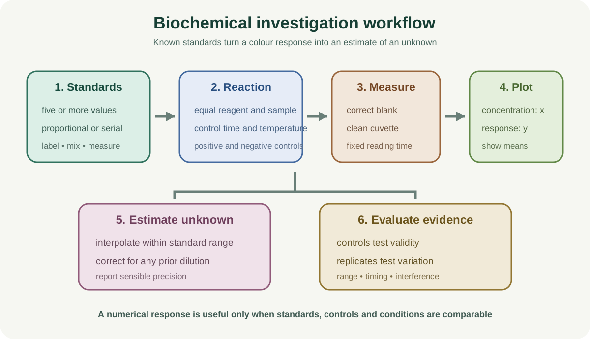
Biochemical Tests, Dilutions and Colorimetry develops Paper 3 and Paper 5 skills through controlled tests, quantitative concentration series, calibration curves and defensible identification of unknowns. The molecular basis of carbohydrates, proteins and lipids belongs in the theory hub. This practical note owns preparation, measurement, safety, records, inference and evaluation.
1. Build a valid comparison
Identify the independent variable, commonly concentration or sample identity, and a measurable dependent variable such as absorbance, transmission, time to a colour endpoint or final colour category. Standardise total volume, reagent concentration and volume, temperature, reaction time, pH, mixing and sample preparation.
Use enough independent-variable values to establish a pattern. Cambridge expects a minimum of five in practical decision-making. Replicate each value so variation can be assessed and a mean calculated. Random variation between repeats is evidence to report, not a reason to select the most convenient result.
A control reveals what happens without the tested component. A negative control might replace sample with distilled water. A positive control contains a known target and confirms that reagents and conditions can produce the expected response.
2. Prepare proportional dilutions
A proportional dilution mixes stock solution and diluent directly to reach each target concentration. The stock volume multiplied by stock concentration equals final volume multiplied by target concentration.
For a fixed final volume, calculate each stock volume, then add diluent until the final volume is reached. For example, to prepare 10 cubic centimetres of 40 percent solution from a 100 percent stock, use 4 cubic centimetres of stock and 6 cubic centimetres of diluent.
Prepare each concentration independently with clean equipment. Label tubes before transferring liquid. Use suitable syringes or pipettes and read scales at eye level. Small volumes can carry large percentage uncertainty, so choose stock and final volumes that the available apparatus can measure reliably.
3. Prepare serial dilutions
A serial dilution transfers a fixed proportion from one tube into a fixed volume of diluent in the next. If equal volumes are mixed at every step, each concentration is half the previous one. Other dilution factors follow from the volumes used.
Mix each tube thoroughly before transferring to the next, use a clean tip or rinsed transfer device and keep transfer volumes consistent. A mistake early in the sequence affects all later tubes, so serial dilution is efficient but propagates error.
Write concentrations for every tube before beginning. Remember that transferring liquid onward can change the volume remaining but not the concentration in the source tube.
Check this topic from memory
Attempt the matching topic bank before reopening the notes. Use each missed idea to decide what to review next.
Add Benedict's solution to the sample in a standardised ratio and heat in a controlled hot-water bath. A positive result progresses from blue through green, yellow and orange to brick red as reducing-sugar concentration increases under comparable conditions.
Use identical heating time, temperature and total volume. A naked flame beneath individual tubes gives inconsistent heating and unnecessary risk. Benedict's reagent is an irritant and hot glass or water can burn, so use eye protection, a test-tube holder and controlled heating.
Colour categories give semi-quantitative evidence. A colorimeter or standard colour scale can improve objectivity, but precipitate can scatter light and complicate absorbance readings.
5. Iodine test for starch
Add iodine in potassium iodide solution to the sample. A blue-black colour supports the presence of starch; iodine remains yellow-brown when starch is absent.
Standardise reagent volume, sample volume and observation background. Use a white tile for clearer comparison. This is usually qualitative unless an explicitly calibrated quantitative method is developed.
Iodine solution is an irritant and can stain. Use eye protection, avoid skin contact and clean spills promptly according to laboratory instructions.
6. Biuret test for protein
Under the specified method, Biuret reagent produces a lilac or purple colour when peptide bonds are present and remains blue in a negative result. Use the volumes and reagent order stated in the task because formulations vary.
Standardise mixing and waiting time before comparing colours. Copper-containing and alkaline reagents require eye protection and careful handling. A positive result indicates peptide bonds, not the identity of a particular protein.
Turbid or strongly coloured samples can interfere with visual judgement. A sample blank containing the sample but not the colour-forming reagent can help account for background absorbance in a colorimetric method.
7. Emulsion test for lipids
Mix the sample with ethanol so lipids dissolve, then add the mixture to water. A cloudy white emulsion supports the presence of lipid. The emulsion forms because lipid becomes dispersed as small droplets when transferred into water.
Ethanol is flammable. Keep it away from flames and other ignition sources. Use small volumes, replace stoppers promptly where directed and wear eye protection.
Cloudiness should be compared against a negative control because insoluble sample material can mimic an emulsion. Filter or standardise sample preparation when appropriate.
8. Use a colorimeter correctly
A colorimeter measures light transmitted through, or absorbed by, a sample at a chosen wavelength or filter. Select a filter that gives strong discrimination for the coloured product. More colour commonly gives greater absorbance and lower transmission, but use the relationship stated or established in the experiment.
Set the instrument with a blank containing all components except the substance responsible for the measured colour. Fill clean cuvettes consistently, remove bubbles, wipe clear faces and orient cuvettes the same way. Fingerprints, scratches, differing volumes and suspended particles alter light transmission.
Measure standards and unknowns under the same reaction time and temperature. If colour continues developing, readings taken at different times are not comparable.
9. Construct and use a calibration curve
Prepare standards of known concentration covering the likely unknown. Plot concentration on the x-axis and absorbance or transmission on the y-axis. Include means when replicates are available and draw the appropriate line or curve from the pattern.
Read an unknown by moving from its instrument value to the calibration line, then down to the concentration axis. Interpolation within the standards is more defensible than extrapolation beyond them. If the unknown lies outside the range, dilute it by a known factor, remeasure and correct the final concentration.
A calibration curve estimates concentration only under the same conditions. It does not compensate for an interfering coloured compound or a reaction that differs between standard and sample.
10. Record qualitative and quantitative evidence
Raw values go in a table with descriptive headings and units in headings. Use consistent precision for measurements from the same instrument. Record colours with specific terms rather than “changed” or “positive.”
When ranking colour by eye, define the scale before collecting data. A numbered category is ordinal, not automatically continuous. Do not calculate a biologically meaningful mean from arbitrary categories unless the question and design justify it.
Photographs can support a record only if lighting, background, camera settings and timing are standardised. They do not automatically remove observer bias.
11. Evaluate biochemical evidence
Separate accuracy from repeatability. Closely grouped colorimeter readings may be repeatable but inaccurate if the blank was wrong. Replicates expose random variation; standards and appropriate controls test validity.
An unknown that falls between calibration values should be reported as an estimate with sensible precision. A saturated instrument reading requires dilution or a different range, not a forced extrapolation.
Match each improvement to a cause: use a thermostatically controlled water bath to reduce temperature variation, timed reagent addition to equalise reaction duration, volumetric equipment to reduce dilution error, or an instrument and calibration curve to reduce subjective colour matching.
Worked application: prepare and use a glucose calibration
Prepare five glucose standards from a 10 grams per cubic decimetre stock, each with a final volume of 10 cubic centimetres. For a 6 grams per cubic decimetre standard, use 6 cubic centimetres of stock and 4 cubic centimetres of water. Treat every standard and the unknown with equal Benedict's reagent, heating time and temperature. After a validated colour measurement, plot mean response against concentration. If the unknown response lies between the 4 and 6 standards, interpolate from the curve. If the sample was diluted twofold before testing, multiply the interpolated concentration by two and report the uncertainty created by the curve and repeats.
Common misconceptions and corrections
Calling every dilution serial. Proportional dilutions can be made independently from stock.
Adding the same water volume to every stock volume. Final concentration depends on both volumes.
Forgetting to mix before a serial transfer. The next tube then receives the wrong concentration.
Assuming serial dilution errors affect one tube only. Early errors propagate.
Using fewer than five concentrations without justification. A useful trend needs a suitable range and intervals.
Calling distilled water a positive control. It is commonly a negative control.
Omitting a known positive. Reagent failure may then be mistaken for an absent molecule.
Heating Benedict's tubes directly over flames. Use a controlled water bath.
Calling every green Benedict's result negative. It can indicate a low reducing-sugar concentration.
Saying iodine identifies glucose. It tests for starch.
Saying Biuret identifies one named protein. It detects peptide bonds.
Calling insoluble debris a lipid emulsion. Compare with a negative control.
Using ethanol near a flame. Ethanol is flammable.
Zeroing a colorimeter with plain water regardless of method. The blank should contain relevant background components.
Holding cuvettes on clear optical faces. Fingerprints alter readings.
Leaving bubbles in a cuvette. They disrupt the light path.
Reading standards at different reaction times. Developing colour becomes incomparable.
Plotting concentration on the y-axis automatically. It is the independent variable and belongs on x.
Extrapolating far beyond standards. Dilute the unknown into the calibrated range.
Treating repeatability as proof of accuracy. A systematic error can produce close wrong values.
Reporting a long calculator display. Precision must reflect apparatus and calibration.
Assessment guidance
A strong method states exact volumes, concentrations, apparatus, order, timing, temperature and mixing. Distinguish proportional from serial dilution and show how each target is obtained. Name both positive and negative controls where they test different failure modes. For biochemical tests, state the reagent, controlled treatment and expected observation without inventing quantitative certainty. Colorimetry answers need the correct blank, consistent cuvette handling, standards spanning the unknown and interpolation from a labelled calibration curve. Evaluation should connect subjective endpoints, temperature drift, timing or dilution uncertainty to a specific consequence and improvement. Safety marks require hazard, risk and precaution, not “be careful.”
Retrieval practice
Prepare five proportional concentrations and a five-step serial dilution on paper. Match Benedict's, iodine, Biuret and emulsion tests to reagent, treatment, positive observation and hazard. Design positive and negative controls for three unknowns. Reconstruct a colorimeter checklist, draw a calibration curve and process one diluted unknown. Finish by converting five vague errors into cause-effect-improvement chains and separating repeatability from accuracy.
Cambridge International, Biology 9700 syllabus for examinations in 2025, 2026 and 2027, Practical Assessment sections for Paper 3 and Paper 5 decisions, dilution, measurement, unknown identification, risk, presentation, analysis and evaluation.