

Cambridge IGCSE Biology Notes 2: Organisation of the Organism

Study guide

Cambridge IGCSE Biology 0610 and 0970 notes on plant, animal and bacterial cells, organelle functions, specialised cells, levels of organisation, magnification and size conversion.

Topic 2 of Cambridge IGCSE Biology 0610 and 0970 connects cell structures to their functions, then builds from specialised cells to tissues, organs, organ systems and organisms. It also requires magnification and size calculations. Core candidates work in millimetres; Extended candidates also convert between millimetres and micrometres.
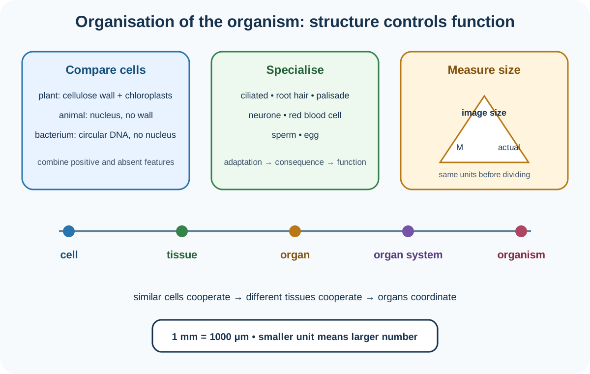
A Cambridge IGCSE Biology organisation map comparing plant, animal and bacterial cells, linking specialised cells to levels of organisation and connecting image size, magnification and actual size

2.1 Cell structure

Structures shared by plant and animal cells

The cell membrane controls movement of substances into and out of the cell. It is a boundary with selective transport roles, not a rigid supporting wall.

Cytoplasm is where many chemical reactions occur. It contains enzymes and surrounds the cell structures.

The nucleus contains genetic material and controls cell activities by controlling which proteins are produced.

Ribosomes are the sites of protein synthesis. Mitochondria are the sites of aerobic respiration, where energy is released from nutrient molecules for cellular processes.

Plant-cell structures

The cellulose cell wall supports the cell, helps maintain shape and prevents bursting when water enters. It is freely permeable and lies outside the cell membrane.

Chloroplasts contain chlorophyll, which absorbs light energy for photosynthesis. They occur in photosynthetic plant cells but are not present in every plant cell, for example most root cells.

A large permanent vacuole contains cell sap, a solution of sugars and salts. Water entering the vacuole contributes to turgor pressure against the cell wall and helps support non-woody tissues.

Plant cells also have a cell membrane, cytoplasm, nucleus, ribosomes and mitochondria.

Animal cells

Animal cells have a cell membrane, cytoplasm, nucleus, ribosomes and mitochondria. They do not have a cellulose cell wall or chloroplasts. Small temporary vacuoles may occur, but the large permanent cell-sap vacuole is a characteristic plant-cell feature in this comparison.

The absence of a wall allows varied cell shapes and movement, but support and shape can still come from the cytoskeleton, surrounding tissues or extracellular material beyond this syllabus detail.

Bacterial cells

A bacterial cell has a cell wall, cell membrane, cytoplasm and ribosomes. Its genetic material includes a circular DNA molecule and may include plasmids, which are smaller DNA loops.

Bacteria have no nucleus. Their circular DNA lies free in the cytoplasm. They also lack mitochondria and chloroplasts; relevant reactions occur using the cell membrane and cytoplasm.

The bacterial wall is not described as cellulose in this syllabus boundary. Do not transfer the composition of plant walls to all cell walls.

Compare by evidence

Use positive and negative features together. A cell wall and chloroplasts support identification as a photosynthetic plant cell. A cell wall with circular DNA but no nucleus supports a bacterium. A nucleus without a wall or chloroplasts supports an animal cell.

One incomplete image may not show every structure. Identify only what the image and question justify, then use the expected cell-type comparison carefully.

New cells

New cells are produced by division of existing cells. Cells do not assemble spontaneously from non-living material. The detailed processes of mitosis and meiosis belong to later syllabus topics.

Specialised cells and their functions

Specialisation means that a cell has structures suited to a particular function.

Ciliated cells

Ciliated cells move mucus in the trachea and bronchi. Beating cilia sweep mucus containing trapped particles and microorganisms toward the throat. The mucus is produced by nearby goblet cells, not by the cilia themselves.

Root hair cells

Root hair cells absorb water and mineral ions. Their long projection provides a large surface area in contact with soil water. A thin wall gives a short movement distance, and mitochondria supply energy for active transport of mineral ions.

Palisade mesophyll cells

Palisade mesophyll cells carry out photosynthesis. They contain many chloroplasts and are positioned near the upper leaf surface where light is strong. Their elongated arrangement helps pack many photosynthetic cells into the tissue.

Neurones

Neurones conduct electrical impulses. Their long extensions carry signals over distance, while branched endings connect with other cells. The detailed synapse mechanism belongs to Topic 14.

Red blood cells

Red blood cells transport oxygen. Their biconcave shape gives a large surface area and short diffusion distance, and they contain haemoglobin. Mammalian red blood cells lack a nucleus, leaving more space for haemoglobin, and are flexible enough to pass through capillaries.

Sperm and egg cells

Sperm and egg cells are gametes used in reproduction. A sperm cell has a flagellum for movement, many mitochondria to supply energy and an acrosome containing enzymes involved in penetrating egg coverings. Its haploid nucleus carries genetic information.

An egg cell is large and contains cytoplasm with nutrient reserves for early development. Its haploid nucleus carries genetic information, and its cell-surface changes after fertilisation help prevent entry of additional sperm.

State the syllabus-level function before adding an adaptation. A long shape alone does not prove a cell is a neurone, and many mitochondria support any process requiring substantial energy.

Levels of organisation

A cell is the basic unit of living organisms. A tissue is a group of similar cells working together for a shared function. An organ contains different tissues working together. An organ system contains organs coordinating a major body function. An organism is the complete living individual.

For example:

muscle cellmuscle tissueheartcirculatory systemhuman. \text{muscle cell}\rightarrow\text{muscle tissue}\rightarrow\text{heart}\rightarrow\text{circulatory system}\rightarrow\text{human}.

The heart is an organ because muscle, nervous, connective and other tissues cooperate. The circulatory system includes the heart and blood vessels working together in transport.

In a plant, a palisade cell contributes to palisade mesophyll tissue, which contributes to the leaf organ, which works with roots and stems within the plant's transport and nutrition systems.

2.2 Size of specimens

Magnification compares image size with actual size:

M=IA. M=\frac{I}{A}.

Here M is magnification, I is image size and A is actual size.

Rearrange as needed:

A=IM. A=\frac{I}{M}.

Magnification has no unit because it is a ratio of two lengths in the same unit. Convert image and actual size to matching units before dividing. A scale bar provides its own route: compare measured image length with measured scale-bar length, then multiply by the real scale-bar value.

Core calculations use millimetres. Extended candidates also use

1,mm=1000,μm. 1,\pu{mm}=1000,\pu{\mu m}.

Multiply millimetres by 1000 to obtain micrometres. Divide micrometres by 1000 to obtain millimetres. A micrometre is smaller, so the numerical value in micrometres is larger for the same length.

Magnification is not resolution. Magnification makes an image larger; resolution is the ability to distinguish close points as separate.

Worked application: identify and size a cell

An image shows a cell with a wall, membrane, cytoplasm, ribosomes and circular DNA but no nucleus. These combined features identify a bacterial cell, not a plant cell, because plant cells have a nucleus and their wall alone is not decisive. The image length is 48,mm48,\pu{mm} at magnification ×12,000\times 12,000. Actual length is 48/12,000=0.0040,mm48/12,000=0.0040,\pu{mm}. Extended conversion gives

Common misconceptions and corrections

  • Calling the cell membrane a supporting wall. It controls exchange; the plant wall gives rigid support.
  • Saying cytoplasm is empty fluid. Many enzyme-controlled reactions occur there.
  • Saying the nucleus makes energy. Mitochondria are the aerobic-respiration sites.
  • Saying mitochondria make protein. Ribosomes synthesise proteins.
  • Giving every plant cell chloroplasts. Non-photosynthetic cells such as root cells usually lack them.
  • Putting the cell membrane outside the cell wall. The wall lies outside the membrane.
  • Calling cell sap cytoplasm. Cell sap is inside the vacuole.
  • Giving animal cells cellulose walls. They have no cellulose cell wall.
  • Calling a bacterium's DNA a nucleus. Its circular DNA lies free in cytoplasm.
  • Giving bacteria mitochondria. They have no mitochondria.
  • Calling all cell walls cellulose. That description applies to plant walls here.
  • Identifying a cell from one feature only. Combine nucleus, wall, chloroplast and DNA evidence.
  • Saying new cells form from nutrients directly. They arise by division of existing cells.
  • Saying cilia produce mucus. Cilia move mucus.
  • Giving a root hair cell a long root. The cell has a surface projection.
  • Saying root hairs absorb only water. They also absorb mineral ions.
  • Giving palisade cells few chloroplasts. Their photosynthetic role is supported by many chloroplasts.
  • Saying neurones carry blood. They conduct electrical impulses.
  • Giving red blood cells a large nucleus. Mammalian red blood cells lack one.
  • Calling the sperm flagellum a cilium. It is a flagellum used for movement.
  • Saying the egg swims toward sperm. Sperm are motile; the egg's large cytoplasm supports early development.
  • Calling a tissue a group of organs. A tissue is a group of similar cells.
  • Calling the heart an organ system. It is one organ within the circulatory system.
  • Using image and actual lengths in different units. Convert before calculating.

Assessment guidance

Identify cell type from a combination of visible structures and state each structure's function precisely. For specialised cells, link one named adaptation to the exact syllabus function rather than listing features without consequences. Keep cell, tissue, organ, organ system and organism in order and use an example with genuine cooperation between levels. In size questions, write the magnification equation, convert both lengths to the same unit, substitute and include the final length unit. Extended candidates should check the direction of the millimetre-micrometre conversion and judge whether the result is biologically plausible.

Retrieval practice

Draw a comparison table for plant, animal and bacterial cells, then identify three unknown cells from mixed positive and negative evidence. Link each of the six specialised cells to two useful adaptations, build one animal and one plant organisation chain, and solve magnification, actual-size and millimetre-micrometre conversion problems using both stated magnification and scale bars.

Theory and practical ownership

This theory note owns cell structures, functions, specialisation, biological organisation and magnification relationships. The separate Biology practical hub owns microscope operation, slide preparation, biological drawing, field-of-view measurement and experimental handling of specimens.

Return to the Biology theory hub or use the Biology practical hub for microscopy, drawing and specimen-measurement skills.

Official source

Cambridge International, Biology 0610 syllabus for examinations in 2026, 2027 and 2028 and Biology 0970 syllabus for examinations in 2026, 2027 and 2028, Topic 2 sections 2.1 Cell structure and 2.2 Size of specimens.

Check this topic from memory

Attempt the matching topic bank before reopening the notes. Use each missed idea to decide what to review next.

Start the topic quiz

Sources

  1. Cambridge IGCSE Biology 0610 syllabus for 2026-2028
  2. Cambridge IGCSE (9-1) Biology 0970 syllabus for 2026-2028