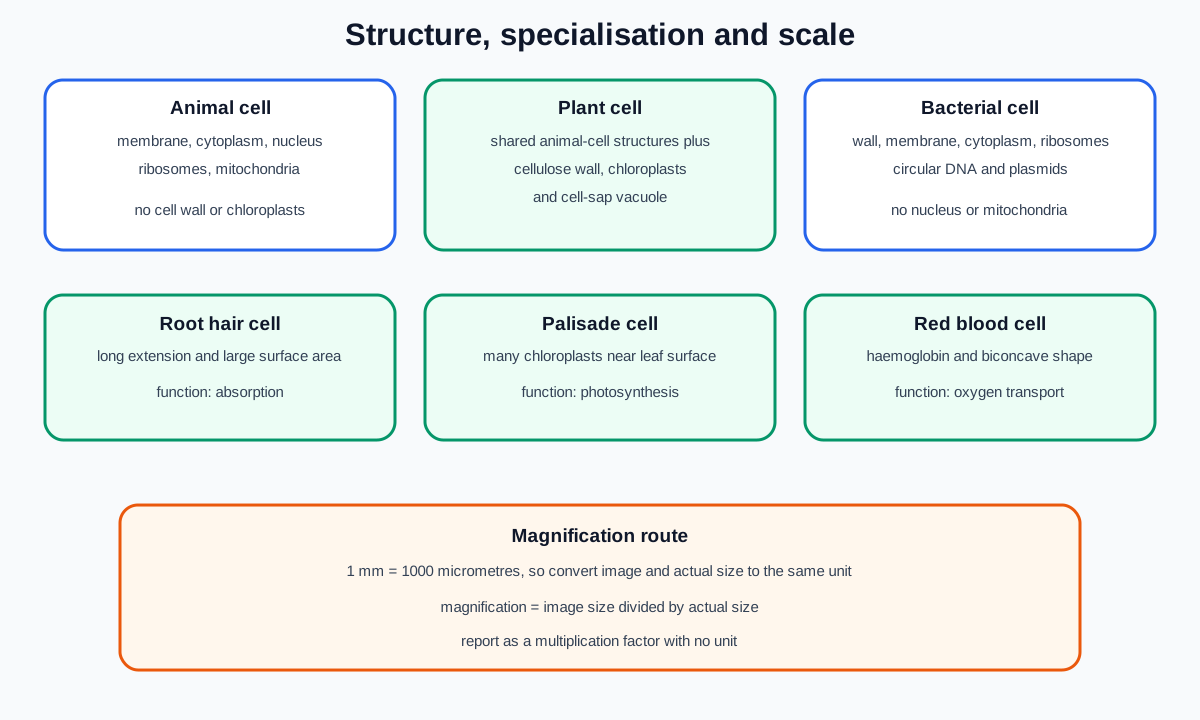
Cambridge IGCSE Combined Science Biology B2 covers plant, animal and bacterial cell structure; the functions of listed plant and animal structures; levels of organisation; three named specialised cells; and magnification calculations using millimetres and micrometres. Keep structure, function and scale as three connected but distinct questions.
Structures shared by plant and animal cells
Plant and animal cells both contain:
a cell membrane, which controls movement of substances into and out of the cell
cytoplasm, where many chemical reactions occur
a nucleus, which contains genetic material and controls cell activities
ribosomes, where proteins are made
mitochondria, where aerobic respiration releases energy
Diagrams are simplified. A typical cell contains many mitochondria and ribosomes, although a two-dimensional drawing may show only a few. The cell membrane is the selectively controlling boundary in both cell types.
Structures characteristic of the syllabus plant-cell model
Plant cells also contain:
a cellulose cell wall, which supports the cell, maintains shape and helps prevent bursting when water enters
chloroplasts, which contain chlorophyll and absorb light for photosynthesis
a vacuole containing cell sap, which contributes to internal support when the cell is turgid
The cell wall is outside the cell membrane. It is freely permeable compared with the membrane, so it does not replace the membrane's role in controlling movement.
Not every plant cell has chloroplasts. Root hair cells usually grow underground and do not photosynthesise, so they are not expected to contain them. “Plant cell” diagrams usually show a photosynthetic example, not every plant cell in every tissue.
Plant cells often appear more regular because the wall resists shape change. Animal cells lack a cell wall and chloroplasts. Animal cells can have small vacuoles, but the large cell-sap vacuole is a feature of the syllabus plant-cell comparison.
Bacterial cell structure
A bacterial cell is limited in this syllabus to:
cell wall
cell membrane
cytoplasm
Check this topic from memory
Attempt the matching topic bank before reopening the notes. Use each missed idea to decide what to review next.
The circular DNA lies free in the cytoplasm rather than inside a nucleus. Plasmids are smaller circular pieces of DNA. A bacterium has no nucleus, mitochondria or chloroplasts in the model required here.
Do not say bacteria have no genetic material merely because they lack a nucleus. Their circular DNA and plasmids are identifiable genetic structures.
The syllabus asks candidates to describe this bacterial structure and identify it in diagrams and images. Do not import unrelated bacterial classifications or disease detail into B2.
Identify cells from combinations of evidence
A cell wall alone does not prove a cell is a plant cell because bacteria also have cell walls. Look for the complete combination.
nucleus plus cell membrane, with no cell wall: animal-cell evidence
nucleus, cellulose wall, vacuole and possibly chloroplasts: plant-cell evidence
circular DNA with no nucleus, plus plasmids: bacterial-cell evidence
Absence from a drawing is not always biological absence. First ask whether the image is complete, labelled and at a scale where the structure should be visible.
In a photomicrograph, identify boundaries and internal regions from evidence rather than expecting the colour scheme used in a textbook diagram.
Link each structure to one precise function
Structure-function questions need a named part and an action. “The nucleus is important” is not enough. State that it contains genetic material and controls cell activities.
Do not swap the roles of ribosomes and mitochondria. Ribosomes make proteins. Mitochondria are the site of aerobic respiration and energy release.
Do not say chloroplasts “make sunlight.” They absorb light energy using chlorophyll and support photosynthesis.
For a cell wall, distinguish support from transport control. For a vacuole, distinguish cell sap and turgor support from the nucleus's control role.
Levels of organisation
A cell is the basic structural unit of a living organism.
A tissue is a group of cells with similar structures working together to perform a shared function. Muscle tissue contains cells adapted for contraction.
An organ is a structure made of different tissues working together for particular functions. The heart contains muscle, nervous and other tissues.
An organ system is a group of organs working together. The circulatory system includes the heart and blood vessels.
An organism is an individual living thing formed from coordinated systems in a multicellular example.
The sequence is:
cell to tissue to organ to organ system to organism.
Size alone does not define the level. An organ is not merely a large tissue; it contains different tissues with coordinated functions.
Specialised cell 1: root hair cell
The syllabus function is absorption.
The long hair-like extension provides a large surface area for uptake of water and mineral ions from soil. A thin boundary gives a short movement distance. The cell is close to xylem tissue, so absorbed materials can enter transport pathways.
Do not claim the extension is a root or that it pushes through soil as locomotion. It is part of one epidermal cell.
Chloroplasts are not an expected adaptation because root hair cells are usually not exposed to light.
Specialised cell 2: palisade mesophyll cell
The syllabus function is photosynthesis.
Palisade cells contain many chloroplasts to absorb light. They are arranged near the upper surface of a leaf, where light intensity is commonly greater. Their elongated arrangement allows many photosynthetic cells to fit into the tissue.
The chloroplast, rather than the whole cell wall or vacuole, directly contains chlorophyll. A palisade cell still has the shared structures needed for respiration, protein synthesis and cellular control.
Specialised cell 3: red blood cell
The syllabus function is transport of oxygen.
Red blood cells contain haemoglobin, which binds oxygen. Their biconcave shape provides a large surface area relative to volume and a short diffusion distance. In mammals, mature red blood cells have no nucleus, leaving more internal space for haemoglobin. Their flexible shape helps them pass through narrow capillaries.
Do not call oxygen transport the job of the cell membrane alone. The whole adaptation set supports loading, carriage and unloading of oxygen.
Magnification compares image and actual size
Magnification is:
M=AI
Here, M is magnification, I is image size and A is actual size.
Rearrange before substituting:
A=MI
I=M×A
Magnification has no unit because image size and actual size are expressed in the same unit before division. Write the answer as, for example, ×400, not 400mm.
Convert millimetres and micrometres first
One millimetre equals 1000 micrometres:
1mm=1000um
To convert millimetres to micrometres, multiply by 1000. To convert micrometres to millimetres, divide by 1000.
Always place image size and actual size in the same unit before using the magnification formula. A correct division with inconsistent units gives a meaningless value.
Measure a printed or displayed image using the unit requested. If the image has been resized, use the scale bar or dimensions in the question rather than an assumed original size.
Worked application: identify and calculate
An image shows a cell with a nucleus, cell wall, large vacuole and many chloroplasts, so the complete evidence supports a plant palisade cell rather than a bacterium, which would lack a nucleus and contain circular DNA. The image length is 48 mm and the actual cell length is 120 micrometres. Convert 48 mm to 48 000 micrometres, then calculate magnification as 48,000/120=400. Report ×400, with no unit. The many chloroplasts and position in palisade tissue support photosynthesis, while the wall alone would not be enough to identify the cell as plant rather than bacterial.
Common misconceptions and corrections
Saying only plant cells have membranes. Both plant and animal cells have cell membranes.
Saying the wall controls entry and exit. The cell membrane performs selective control.
Calling the plant wall a cell membrane. They are separate layers.
Assuming every plant cell contains chloroplasts. Non-photosynthetic cells may not.
Putting chlorophyll in the vacuole. Chlorophyll is in chloroplasts.
Saying mitochondria make proteins. Ribosomes make proteins.
Saying ribosomes release energy by aerobic respiration. Mitochondria do.
Saying bacteria have no DNA. They have circular DNA and may have plasmids.
Drawing bacterial DNA inside a nucleus. Bacteria lack a nucleus.
Identifying a plant cell from a wall alone. Bacteria also have walls.
Calling a tissue a group of organs. A tissue is a group of similar cells working together.
Calling an organ one kind of cell. Different tissues work together in an organ.
Skipping organ system in the hierarchy. Keep the full five-level sequence.
Calling a root hair extension a separate organ. It is part of one cell.
Giving root hair cells chloroplasts as their main adaptation. Their function is absorption underground.
Saying palisade cells only photosynthesise and do not respire. Living cells also respire.
Saying red blood cells carry oxygen because of a nucleus. Mature mammalian cells lack one and contain haemoglobin.
Dividing sizes expressed in different units. Convert first.
Giving magnification in millimetres. It is a ratio with no unit.
Multiplying by magnification when finding actual size. Divide image size by magnification.
Assessment guidance
Cell-identification answers should cite a combination of visible structures rather than one feature. Structure-function questions need the named structure and a precise biological action. For organisation, use examples that contain the correct lower-level components and preserve the cell-to-organism order. Specialised-cell questions should link a structural feature to the exact syllabus function, not list unrelated facts. In magnification calculations, write the formula, convert millimetres and micrometres to a common unit, substitute clearly and report magnification with a multiplication sign but no unit. Check whether a scale bar remains valid if an image is resized.
Retrieval practice
Draw unlabelled plant, animal and bacterial cells, then add only the official structures. Build a structure-function matching grid and identify cells from twenty incomplete images. Reconstruct the five levels of organisation with two examples each. Explain three adaptation chains for every specialised cell. Complete thirty magnification problems that mix millimetres, micrometres, image size, actual size and scale bars.
Topic ownership
This note owns cell structure, listed functions, organisation, the three named specialised cells and magnification. B3 owns membrane transport mechanisms, B6 and B7 own nutrition, and B12 owns respiration equations. The practical hub owns microscope handling, specimen measurement and experimental evaluation.