Cambridge IGCSE Combined Science Biology B9 develops the circulatory system as a pump, vessels and valves; the structures and action of the mammalian heart; exercise and heart rate; coronary heart disease; arteries, veins and capillaries; and the identification and functions of blood components.
The circulatory system is a one-way transport system
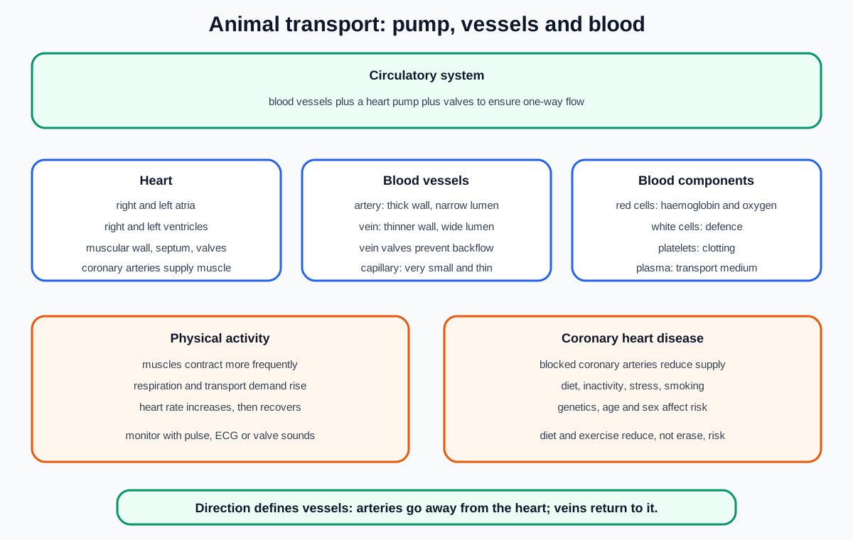
The circulatory system is a system of blood vessels with a pump and valves to ensure one-way flow of blood.
The heart is the pump. Blood vessels form routes through the body. Valves prevent backflow so pressure changes move blood in the intended direction.
This definition is structural as well as functional. “Blood moving around the body” omits the required pump, vessels and one-way control.
Blood supplies substances to tissues and removes substances from them. The exact transport roles of plasma and red blood cells are developed later in this topic.
Identify the four heart chambers
The mammalian heart contains a right atrium, right ventricle, left atrium and left ventricle.
The atria are the upper receiving chambers. The ventricles are the lower pumping chambers. In many front-view diagrams, the anatomical left side appears on the right side of the page, so use labels and vessel connections rather than assuming page direction.
The chamber walls are muscular because contraction produces pressure. Ventricular walls are thicker than atrial walls because ventricles pump blood away from the heart, while atria move blood a shorter distance into ventricles.
Within this Combined Science boundary, identify the chambers and understand their contraction sequence. Detailed named-vessel circuit tracing is not a listed B9 requirement.
The septum separates the two sides
The septum is the wall separating the left and right sides of the heart.
It prevents blood on the two sides from mixing directly inside the heart. This separation supports different pressure conditions and destinations for blood leaving each side.
Do not call the septum a valve. It is a dividing wall. Valves have a different role: preventing backflow between connected regions.
In a diagram, trace the boundary running between the atria and between the ventricles rather than labelling an outer heart wall as the septum.
One-way valves respond to pressure differences
Valves ensure one-way blood flow by opening when pressure behind them is greater and closing when pressure would otherwise drive blood backward.
Valves do not actively pump blood and they do not decide when to open. Their movement follows pressure differences created by heart-muscle contraction and relaxation.
Check this topic from memory
Attempt the matching topic bank before reopening the notes. Use each missed idea to decide what to review next.
The syllabus requires one-way valves as identifiable heart structures and their action during heart function. A good answer names the backflow that is prevented rather than saying only that valves “control blood”.
Sounds heard through a stethoscope are associated with valves closing. Listening to these sounds is one of the specified ways of monitoring heart activity.
Coronary arteries supply the heart muscle
Coronary arteries are blood vessels on the heart surface that supply the heart muscle with oxygen and nutrients.
The heart is a living, respiring organ. Its muscle needs a continuous supply so it can contract. Blood inside the chambers does not simply provide enough oxygen by diffusing through the thick heart wall.
In diagrams, distinguish coronary arteries on the heart surface from the muscular wall and internal chambers.
Blockage of coronary arteries reduces the blood supply to heart muscle and is central to coronary heart disease.
Blood leaves in arteries and returns in veins
Blood is pumped away from the heart in arteries and returns to the heart in veins.
Direction relative to the heart defines these vessel types. Oxygen content does not define them.
This rule prevents a common error: it remains valid even when the oxygen content of blood differs between particular vessels. For this Combined Science topic, use the direction rule rather than importing unlisted named-vessel routes.
Valves within the heart and valves in veins both prevent backflow, but they occur in different parts of the system.
Atrial and ventricular contraction creates the pumping cycle
The heart functions through coordinated contraction and relaxation of cardiac muscle.
When the atria contract, pressure rises and blood moves into the ventricles through open one-way valves. When the ventricles contract, ventricular pressure rises. The valves between atria and ventricles close to prevent backflow, while blood is driven away from the heart.
When chambers relax, pressure falls and the chambers refill. The valve sequence preserves one-way flow through repeated cycles.
Do not say all four chambers contract together. The atria contract before the ventricles. Also avoid saying contraction “sucks” blood out of the heart; contraction raises pressure and pushes blood.
Monitor heart activity in three specified ways
An ECG, or electrocardiogram, records electrical activity associated with the heart cycle. It provides a pattern over time rather than a direct picture of blood flowing.
Pulse rate can be measured where pressure waves in an artery are detectable. Count beats for a known interval and express the result as beats per minute. A short count can be scaled to one minute, but a longer interval usually reduces the proportional effect of a one-beat counting error.
Heart sounds can be heard with a stethoscope. They are associated with valves closing.
These methods provide different evidence. A pulse is a pressure wave, an ECG records electrical activity, and a stethoscope detects sounds. Do not present them as identical measurements.
Investigate physical activity and heart rate
Measure a resting pulse after the participant has sat quietly. Ask the participant to perform a standardised activity for a fixed time, then measure pulse rate immediately afterward. Continue measuring at regular intervals during recovery.
Keep the activity type, duration, pace, measurement method and recovery intervals consistent. If comparing people, biological differences make perfect control impossible, so use repeated trials and interpret person-to-person variation cautiously.
Record heart rate in beats per minute. Plot heart rate against time, including the resting value, post-exercise value and recovery measurements.
Safety and ethics matter. Screen for known health concerns, use a moderate activity appropriate to the participant, provide space and stable footwear, stop if the participant feels unwell, obtain agreement and avoid public identification of personal results.
Explain why physical activity raises heart rate
Active muscles contract more frequently and need more energy. The rate of respiration increases to transfer this energy.
Blood must deliver oxygen and glucose more rapidly to the muscles and remove carbon dioxide more rapidly. An increased heart rate pumps blood through the circulation more frequently, helping meet these increased transport demands.
After activity stops, heart rate usually returns gradually toward its resting value as demand falls and recovery processes continue.
Do not say the heart rate rises merely because “the body gets tired”. Link muscle activity, respiration, substance transport and pumping rate.
Coronary heart disease can be described in terms of blockage of coronary arteries.
Narrowing or blockage reduces blood flow to part of the heart muscle. Less oxygen and fewer respiratory substrates reach those cells, so aerobic respiration and muscle function can be impaired. A severe blockage can damage heart tissue.
The required description centres on coronary arteries, not valves or the chambers becoming filled with fat.
Coronary heart disease has multiple interacting risk factors. A risk factor changes probability; it does not guarantee that an individual will or will not develop disease.
Evaluate the named risk factors
The specified possible risk factors are diet, lack of exercise, stress, smoking, genetic predisposition, age and sex.
A diet high in particular fats and excessive energy intake can contribute to conditions associated with coronary risk. Lack of exercise can reduce cardiovascular fitness and contribute to unhealthy body mass. Smoking exposes the circulatory system to harmful substances and increases risk. Long-term stress can influence behaviour and physiology.
Genetic predisposition means inherited variation can affect risk. Age and sex are also associated with different risk patterns. These factors cannot be interpreted as personal certainty.
Avoid moral judgement. Some factors can be modified, while others cannot. Population associations also do not prove that one factor alone caused one person's disease.
Diet and exercise can reduce risk
A balanced diet can help manage energy intake and reduce excessive intake of foods associated with increased coronary risk. It should be described as an overall pattern rather than a single “superfood” cure.
Regular suitable exercise can improve cardiovascular fitness and help maintain a healthy body mass. It can also replace some sedentary time.
Diet and exercise reduce risk but cannot remove all risk because smoking, stress, genetics, age, sex and other interacting conditions still matter. Recommendations should be realistic, gradual and appropriate to the individual.
When discussing a strategy, state how it changes a named risk factor and acknowledge limitations rather than promising prevention.
Compare artery, vein and capillary structure
Arteries have relatively thick walls and relatively narrow lumens. They do not require the vein-valve feature in the specified comparison.
Veins have relatively thinner walls, wider lumens and valves.
Capillaries are very small vessels with very thin walls and narrow lumens. At this level, use their small scale when recognising them in diagrams, while keeping the official comparison focused on relative wall thickness, lumen diameter and valves in veins.
Compare like with like. “An artery has a thick wall” is useful only when the reference is clear. A table should state relative thickness, relative lumen width and valve presence.
Relate artery and vein structure to pressure
Arteries transport blood away from the heart at relatively high pressure. Their thick walls withstand this pressure and help maintain the vessel.
Veins return blood at lower pressure. Their walls can be thinner, and their wider lumens reduce resistance to flow. Valves prevent backflow where pressure is low.
Do not explain thick artery walls as allowing diffusion. Transport rather than exchange is their main role. Do not say veins have no pressure at all; the pressure is lower than in arteries.
The syllabus requires pressure relationships for arteries and veins. Avoid importing more detailed wall-layer histology as if it were required.
Blood contains cells, cell fragments and plasma
The four named blood components are red blood cells, white blood cells, platelets and plasma.
Plasma is the liquid in which cells and platelets are carried. A blood sample drawing may show many red blood cells, fewer larger white blood cells and much smaller platelets.
Identify red blood cells by their repeated small disc-like appearance and lack of a visible nucleus in typical mammalian diagrams. White blood cells are larger, less numerous and have a visible nucleus in common photomicrographs.
Platelets are small cell fragments. Detailed clotting stages are not required.
Red blood cells transport oxygen
Red blood cells contain haemoglobin. Haemoglobin binds oxygen and enables red blood cells to transport it.
Their shape gives a large surface area relative to volume, and lacking a nucleus leaves more space for haemoglobin, although the named mark-bearing function is oxygen transport including haemoglobin's role.
Do not say haemoglobin transports the red blood cell. The cell is carried in blood; haemoglobin inside it carries oxygen.
Red blood cells are not the main cells responsible for antibody production or phagocytosis.
White blood cells defend by two named routes
White blood cells defend against pathogens through phagocytosis and antibody production.
In phagocytosis, a white blood cell surrounds and digests a pathogen. Other white blood cells produce antibodies that bind specifically to target antigens.
The syllabus combines both functions under white blood cells. Do not claim every individual white blood cell performs both processes in exactly the same way.
Keep pathogen defence distinct from blood clotting, which is the role of platelets.
Platelets support clotting
Platelets help blood clot. Details of the clotting cascade are not required.
Clotting prevents excessive blood loss from damaged vessels. It also helps prevent pathogens entering through the wound.
A clot is not an antibody response and platelets do not engulf pathogens. Their protective effect comes from sealing the break.
Do not equate clotting with blood becoming solid throughout a vessel. It is a local response to damage under normal circumstances.
Blood cells and platelets are carried suspended in plasma. Dissolved ions and nutrients move between exchange surfaces and tissues. Urea is carried away from tissues for removal. Hormones travel from endocrine organs to target tissues. Much carbon dioxide is transported in the plasma.
Use the full named list when a question asks broadly for plasma functions. Do not say plasma carries oxygen only; oxygen transport is principally linked to haemoglobin in red blood cells.
Worked application: explain an exercise data set
A student's resting heart rate is measured for one minute, then again immediately after two minutes of standardised stepping and at one-minute recovery intervals. The rate rises sharply after exercise and then falls toward the resting value. The rise is explained by more frequent muscle contraction, increased respiration, greater oxygen and glucose demand, and faster carbon-dioxide removal. Repeating the same protocol on another day tests consistency. The student's data cannot by itself prove general fitness differences or coronary risk because one participant, effort variation and biological fluctuation limit inference. A safe conclusion describes this participant's heart-rate response under the tested conditions.
Common misconceptions and corrections
Defining circulation as blood simply moving. Include vessels, a pump and valves for one-way flow.
Calling the septum a valve. It is the wall separating the heart's sides.
Assuming page left is anatomical left. Use labels and structural connections.
Saying all chambers contract together. Atrial contraction precedes ventricular contraction.
Saying arteries always carry oxygenated blood. They are defined by flow away from the heart.
Saying veins always carry deoxygenated blood. They are defined by return to the heart.
Saying coronary arteries carry blood into a chamber. They supply the heart muscle.
Calling a pulse a moving lump of blood. It is a pressure wave in an artery.
Saying an ECG directly shows blood flow. It records electrical activity.
Saying heart sounds are chambers opening. They are associated with valves closing.
Explaining exercise response as tiredness only. Link respiration, transport demand and pumping.
Using unsafe maximal exercise for a class test. Standardise a suitable moderate activity and apply stop rules.
Claiming one exercise result proves fitness. Repeat and limit the conclusion to the evidence.
Describing coronary disease as blocked heart valves. It involves blockage of coronary arteries.
Treating a risk factor as a guaranteed cause. It changes probability.
Claiming diet and exercise remove all risk. Non-modifiable and interacting factors remain.
Using moral judgement about disease. Discuss mechanisms and probabilities.
Saying veins have no pressure. Their pressure is lower than arterial pressure.
Saying artery walls are thick for diffusion. They withstand relatively high pressure.
Forgetting vein valves. They prevent backflow at lower pressure.
Saying plasma is a blood cell. It is the liquid transport medium.
Saying haemoglobin is a white-cell antibody. It carries oxygen in red blood cells.
Saying platelets produce antibodies. They support clotting.
Giving detailed clotting chemistry as required. Clotting details are explicitly not required.
Forgetting the second role of clotting. It prevents blood loss and pathogen entry.
Listing only nutrients in plasma. Include cells, ions, nutrients, urea, hormones and carbon dioxide.
Assessment guidance
Begin circulation answers with pump, vessels, valves and one-way flow. In heart diagrams identify the four chambers, muscular wall, septum, one-way valves and coronary arteries before explaining the atrial and ventricular sequence. Keep ECG, pulse and valve sounds as distinct monitoring evidence. Exercise explanations need muscle respiration, delivery and removal demands, and increased pumping. Coronary answers require artery blockage, cautious treatment of every named risk factor, and mechanism-based diet and exercise discussion. Vessel comparisons need relative wall, lumen and valve features linked to pressure. Blood questions need identification evidence and exact functions for red cells, white cells, platelets and plasma.
Retrieval practice
Label twenty rotated heart diagrams and narrate valve action through one cycle. Design an ethical exercise investigation and interpret a recovery graph. Sort the seven coronary risk factors into modifiable and non-modifiable groups, then explain why the categories do not predict certainty. Rebuild an artery, vein and capillary comparison from memory. Identify cells in varied photomicrographs and write the complete red-cell, white-cell, platelet and plasma function set.
Topic ownership
This note owns the B9 circulatory-system definition, named heart structures and action, three monitoring methods, exercise and heart rate, coronary heart disease, vessel comparison and the full blood-component list. B10 owns transmissible disease and immunity. B13 owns respiration. The dedicated practical hub owns extended data-processing and investigation templates. Pulmonary and systemic circuit tracing and named major vessels are not promoted into this Combined Science B9 boundary.