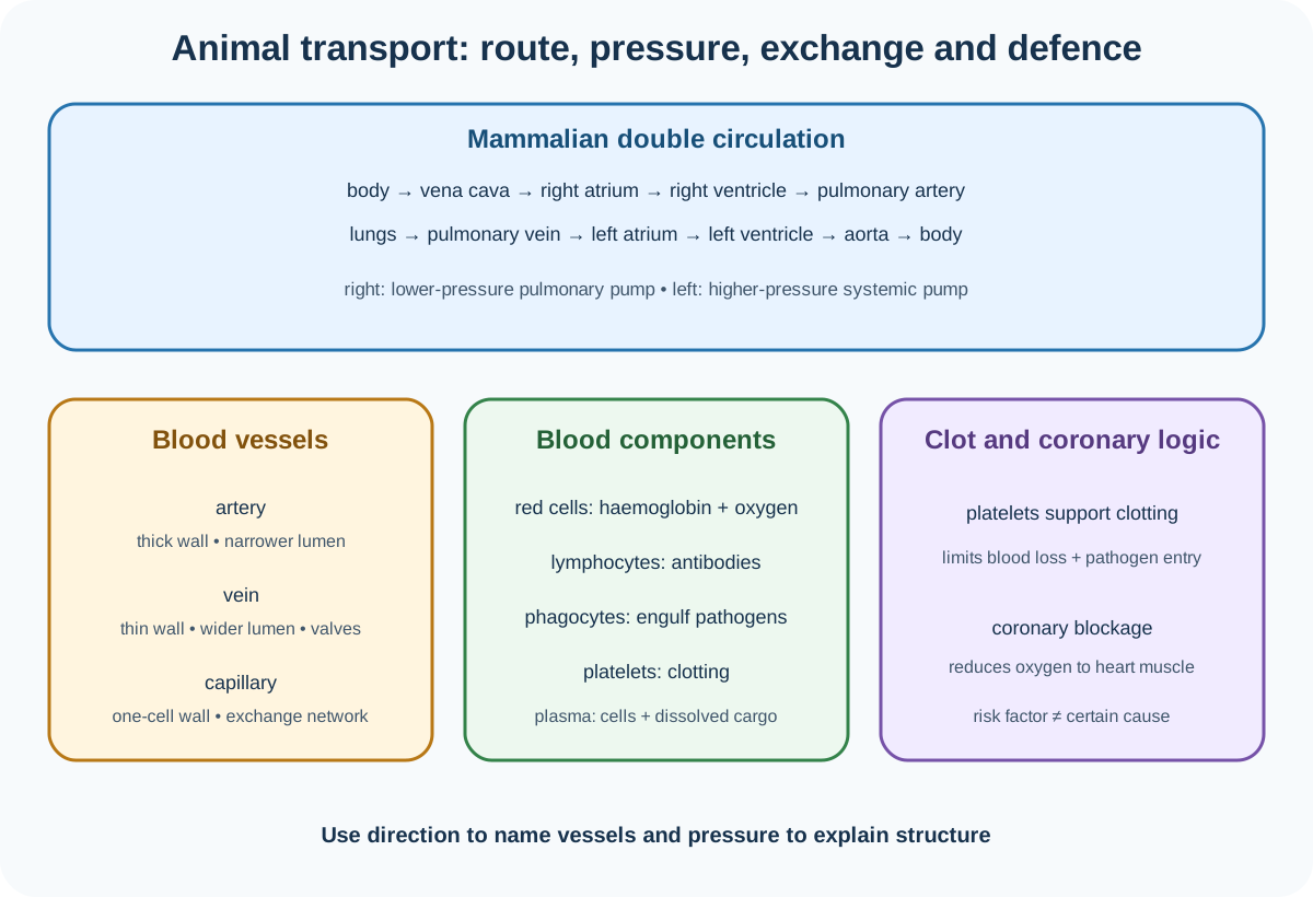
Topic 9 of Cambridge IGCSE Co-ordinated Sciences 0654 and 0973 develops the circulatory system as blood vessels with a pump and valves for one-way flow. Official sections 9.1 to 9.4 cover single and double circulation, mammalian heart structure and function, exercise and coronary risk, vessel-pressure relationships, four named heart-lung vessels, blood cells, plasma and the roles of clotting. Tissue fluid and lymph are not imported into this boundary.
Single and double circulations
A circulatory system contains a pump, blood vessels and valves that ensure one-way blood flow. The heart generates pressure, vessels route blood and valves prevent reversal when pressure changes.
In the single circulation of a fish, blood passes through the heart once in one complete circuit:
heart→gills→body→heart.
Pressure falls as blood passes through gill capillaries before travelling to the rest of the body.
In the double circulation of a mammal, blood passes through the heart twice in a complete journey. The pulmonary circuit carries blood from the right side of the heart to the lungs and back to the left side. The systemic circuit carries blood from the left side to the body and back to the right side.
Check this topic from memory
Attempt the matching topic bank before reopening the notes. Use each missed idea to decide what to review next.
Double circulation allows blood returning from the lungs to be pumped again at high pressure to body tissues, supporting rapid delivery of oxygen and nutrients. The pulmonary circuit can remain at lower pressure, reducing risk of damage to delicate lung capillaries. Separation also prevents oxygenated and deoxygenated blood from mixing in the heart.
The mammalian heart route
Follow the route by vessel, chamber and oxygenation:
Vena cava returns deoxygenated blood from the body to the right atrium.
The right atrium contracts and blood passes through the right atrioventricular valve to the right ventricle.
The right ventricle contracts and sends blood through a semilunar valve into the pulmonary artery.
Pulmonary arteries carry deoxygenated blood to the lungs.
Pulmonary veins return oxygenated blood to the left atrium.
The left atrium contracts and blood passes through the left atrioventricular valve to the left ventricle.
The left ventricle contracts and sends blood through a semilunar valve into the aorta.
The aorta carries oxygenated blood to the body.
Arteries are defined by carrying blood away from the heart and veins by returning blood to it. Oxygen content therefore has exceptions: the pulmonary artery is deoxygenated and the pulmonary vein oxygenated.
The muscular heart wall contracts to generate pressure. Atria have thinner walls because they move blood a short distance into ventricles. The right ventricle pumps to nearby lungs at lower pressure and has a thinner wall than the left ventricle. The left ventricle has the thickest wall because it must generate high pressure for the systemic circuit.
The septum separates the right and left sides, preventing mixing of oxygenated and deoxygenated blood. Atrioventricular valves lie between atria and ventricles. Semilunar valves lie at the exits from ventricles. Valves open when pressure behind them exceeds pressure ahead and close when the pressure difference reverses. Valves do not actively pump.
Coronary arteries supply the heart muscle with oxygenated blood. They are not the vessels carrying blood through the heart chambers.
Monitoring the heart and physical activity
Heart activity can be monitored by an electrocardiogram, or ECG, pulse rate and sounds made as valves close. These measures show different evidence: an ECG records electrical activity, pulse reflects arterial pressure waves and heart sounds arise mainly from valve closure.
During physical activity, muscle cells respire faster. They need more oxygen and glucose and produce carbon dioxide more rapidly. Heart rate rises so blood can deliver substrates and remove carbon dioxide faster. After activity, rate gradually returns toward resting level as demand falls and recovery continues.
An investigation should compare the same person before and after a standardised activity or compare controlled workloads with sufficient recovery and repeats. Method execution and risk control belong to the practical hub.
Coronary heart disease
Coronary heart disease involves blockage or narrowing of coronary arteries, reducing blood and oxygen delivery to heart muscle. Severe restriction can damage muscle and impair pumping.
Cambridge names diet, lack of exercise, stress, smoking, genetic predisposition, age and sex as possible risk factors. A risk factor changes probability; it is not proof that every exposed person will develop disease. Several factors can interact.
A diet that supports healthy body mass and avoids excessive intake of foods associated with raised blood lipids can reduce modifiable risk. Regular exercise can improve cardiovascular fitness and help maintain a healthy mass. These choices reduce risk but do not remove age, sex or inherited predisposition.
Discuss questions require benefits, limitations and interactions. Avoid blaming an individual or claiming certainty from correlation.
Arteries, veins and capillaries
Arteries carry blood away from the heart, usually at high and pulsatile pressure. They have relatively thick muscular and elastic walls and relatively narrow lumens. Thick strong walls withstand pressure; elastic tissue allows stretch and recoil. They do not require valves along most of their length because pressure maintains forward flow.
Veins return blood at lower pressure. They have relatively thinner walls, larger lumens and valves. The large lumen reduces resistance to flow, while valves prevent backflow, especially when surrounding muscles compress veins.
Capillaries connect the arterial and venous sides and provide exchange surfaces. Their walls are one cell thick, giving a short diffusion distance. Their narrow lumen holds red blood cells close to the wall, and extensive networks provide a large total surface area. Slow flow and close tissue contact support exchange of substances.
Named blood vessels
The vena cava returns blood from the body to the heart. The aorta carries blood from the heart to the body. The pulmonary artery travels from heart to lungs and pulmonary vein from lungs to heart.
Use direction and connected organ to identify a vessel. "Artery" or "vein" alone does not state oxygen or nutrient content reliably.
Components of blood
Blood contains red blood cells, white blood cells, platelets and plasma.
Red blood cells transport oxygen using haemoglobin. The 0654 B9 outcome requires this function rather than a list of red-cell structural adaptations.
White blood cells defend against pathogens. Lymphocytes produce antibodies. Phagocytes engulf pathogens by phagocytosis. In photomicrographs, white cells have nuclei and are larger but less numerous than red cells. A lymphocyte commonly has a large round nucleus; a phagocyte may have a lobed nucleus and irregular outline.
Platelets are cell fragments involved in clotting. Plasma is the liquid that transports blood cells, ions, nutrients, urea, hormones and carbon dioxide. Oxygen is mainly carried by haemoglobin rather than simply listed as a principal dissolved plasma cargo in this outcome.
Blood clotting
Clotting prevents excessive blood loss and reduces entry of pathogens through a wound. Platelets participate in clotting, but the reaction stages and named clotting proteins are not required in 0654 B9.
Worked application: trace blood and explain a vessel
An oxygenated red blood cell leaving the lungs enters a pulmonary vein, then the left atrium, left atrioventricular valve, left ventricle, semilunar valve and aorta. After delivering oxygen in body capillaries, it returns through veins and the vena cava to the right atrium. The left ventricle is thicker than the right because systemic flow needs greater pressure over a longer, more resistant circuit. An artery section has a thick wall and narrower lumen to withstand that pressure. At capillaries, a one-cell-thick wall and narrow lumen create a short exchange distance.
Common misconceptions and corrections
Defining circulation without valves. Valves help ensure one-way flow.
Saying fish have double circulation. Their blood passes through the heart once per circuit.
Saying mammalian blood visits the heart once. It returns between pulmonary and systemic circuits.
Claiming double circulation always uses the same pressure. Pulmonary pressure is lower than systemic pressure.
Saying arteries always carry oxygenated blood. The pulmonary artery is deoxygenated.
Saying veins always carry deoxygenated blood. The pulmonary vein is oxygenated.
Sending vena-cava blood to the left atrium. It enters the right atrium.
Sending the pulmonary artery to the body. It travels to the lungs.
Sending the pulmonary vein to the right atrium. It returns to the left atrium.
Calling the aorta a vein. It is the main systemic artery.
Saying atria pump directly around the body. They move blood into ventricles.
Giving both ventricles equal wall thickness. The left is thicker.
Saying the right ventricle is thickest because it pumps deoxygenated blood. Thickness depends on required pressure.
Calling the septum a valve. It separates the two sides.
Calling an ECG a pulse trace. It records electrical activity.
Explaining exercise heart rate only as needing energy. Link respiration, oxygen and glucose delivery, and carbon dioxide removal.
Saying one risk factor guarantees coronary disease. It alters probability.
Treating age and genetic predisposition as lifestyle choices. They are non-modifiable factors.
Claiming exercise removes all risk. It reduces modifiable risk.
Defining an artery by a thick wall only. Direction away from the heart defines it.
Defining a vein by valves only. Direction toward the heart defines it.
Saying arteries have wider lumens than veins. Veins generally have wider lumens relative to wall thickness.
Saying capillaries have thick muscular walls. Their walls are one cell thick.
Saying capillaries only transport blood. They are the main exchange vessels.
Calling all white blood cells antibody producers. Lymphocytes produce antibodies; phagocytes engulf pathogens.
Calling red blood cells the clotting component. Platelets participate in clotting.
Saying plasma transports only water. It carries cells and many dissolved substances.
Giving clotting only a blood-loss role. It also reduces pathogen entry.
Importing tissue-fluid and lymph detail into Topic 9. Those outcomes are not in this official boundary.
Assessment guidance
Trace circulation using connected vessels and chambers rather than memorised oxygen labels. Explain double circulation through pressure, rapid systemic delivery and pulmonary protection. In heart questions, relate wall thickness to distance and pressure, describe contraction in atria then ventricles and explain valves through pressure-driven one-way flow. For vessel comparisons, connect wall, lumen and valves to transported pressure, then give capillary exchange adaptations. Name the four required heart-lung vessels by direction. Separate lymphocyte antibody production from phagocyte engulfment, list every required plasma cargo and state both roles of clotting. Treat coronary factors as risks, not deterministic causes.
Retrieval practice
Draw single and double circulations, then trace blood from vena cava through both circuits to the aorta. Label a heart, compare all chamber walls and predict valve states during contraction. Identify artery, vein, capillary, lymphocyte and phagocyte images, list plasma cargo and explain why clotting limits both blood loss and pathogen entry.
Theory and practical ownership
This theory note owns circulation routes, heart and vessel structure-function relationships, exercise explanations, coronary risk, named vessels, blood-cell identification and clotting. The separate Biology practical hub owns pulse and exercise protocols, recovery periods, participant safety, control variables, repeats, tables, graphs and evaluation.