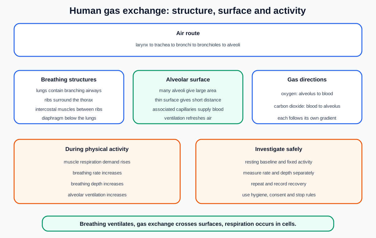
Cambridge IGCSE Co-ordinated Sciences Biology B11 requires confident identification of eleven breathing-system structures, investigation of how physical activity changes breathing rate and depth, and explanation of four human gas-exchange-surface features: large area, thin surface, good blood supply and good ventilation with air.
Keep breathing, gas exchange and respiration distinct
Breathing moves air into and out of the lungs. Gas exchange transfers oxygen and carbon dioxide between air in the alveoli and blood in associated capillaries. Respiration is the set of chemical reactions in cells that releases energy and belongs to B12.
These processes are connected but not interchangeable. Breathing ventilates the gas-exchange surface, blood transports gases, and cells use oxygen in aerobic respiration.
An answer saying “the lungs respire” confuses organ-level gas exchange with cell chemistry. An answer saying “gas exchange is breathing in oxygen” misses transfer across the exchange surface.
This topic does not require the detailed pressure and volume mechanics of inhalation and exhalation. Use the listed structures and exchange features as the assessment boundary.
Identify the lungs
The lungs are paired organs in the thorax. They contain branching air passages ending in many alveoli.
In a front-view diagram, label the lung tissue on both sides of the central trachea and heart region rather than labelling the empty central space.
The lungs provide the organ system in which air is ventilated and gas exchange occurs. They are not hollow bags with one open internal chamber.
When a diagram shows only one side in detail, recognise that bronchioles and alveoli occur throughout lung tissue.
Identify the ribs and intercostal muscles
Ribs form part of the protective framework around the thorax.
Intercostal muscles lie between the ribs. The name itself helps: “intercostal” means between ribs.
In simplified diagrams, rib outlines may appear as curved bands around the lungs, while intercostal muscles occupy spaces between them. Do not label the outer chest line automatically as a muscle.
Their identification matters even though detailed ventilation mechanics are not specified in B11.1.
Identify the diaphragm
The diaphragm is a sheet of muscle below the lungs that separates the thorax from the abdomen.
It is often drawn as a curved dome beneath the lungs. Do not place it above the lungs or inside an alveolus.
Its muscular nature distinguishes it from the air passages. The required task is identification, so use position and shape consistently across unfamiliar diagrams.
Check this topic from memory
Attempt the matching topic bank before reopening the notes. Use each missed idea to decide what to review next.
Avoid saying inhaled air enters the diaphragm. Air remains within the breathing passages and alveoli.
Identify the larynx and trachea
The larynx lies at the top of the trachea and is commonly called the voice box.
The trachea is the main airway leading down from the larynx toward the lungs. It is often shown with supporting rings.
In a label line question, check the endpoint carefully. A line ending at the upper enlarged region may indicate the larynx, while a line ending on the long central tube indicates the trachea.
Do not call the trachea an oesophagus. The oesophagus belongs to the digestive route.
Distinguish bronchi from bronchioles
The trachea divides into two bronchi, with one bronchus entering each lung.
Inside the lungs, the bronchi divide repeatedly into smaller bronchioles.
Use branching order and diameter. A bronchus is one of the large branches directly after the trachea, while bronchioles are the many finer branches deeper within lung tissue.
The plural is bronchi; the singular is bronchus. Do not label every airway branch as a bronchus.
Identify alveoli and associated capillaries
Alveoli are tiny air sacs at the ends of bronchioles. Many alveoli together provide the gas-exchange surface.
Associated capillaries form a blood-vessel network close around the alveoli. In a magnified diagram, separate the air space inside an alveolus from the capillary containing blood cells outside it.
An alveolus is not a blood vessel and a capillary is not an air tube. They are adjacent structures separated by a thin exchange surface.
Singular and plural wording matters: one alveolus, many alveoli.
Trace the air route in order
The required named airway route is:
larynx, trachea, bronchi, bronchioles and alveoli.
The lungs contain the later parts of this route. Ribs, intercostal muscles and diaphragm support breathing but air does not pass through them.
When tracing an image, follow a continuous open airway from the central trachea into increasingly narrow branches. This prevents confusion between a bronchiole and an associated blood vessel.
Do not insert blood capillaries into the air route. They carry blood alongside the alveolar air spaces.
Large surface area increases exchange capacity
Many small alveoli create a very large total surface area.
A larger area allows more oxygen and carbon dioxide particles to cross at the same time, provided the other conditions for exchange are maintained.
The adaptation depends on the total area of many sacs, not on each alveolus being large. A few large sacs would provide less surface area relative to the same volume.
In an explanation, connect feature to consequence: many alveoli give a large area, so more diffusion can occur simultaneously.
A thin surface gives a short diffusion distance
The alveolar exchange surface is thin.
A thin barrier creates a short distance between alveolar air and capillary blood. Particles cross this distance more quickly than they would cross a thick barrier.
Do not say the surface has no wall. A barrier is present, but it is thin.
Avoid using “thin” without its consequence. The assessment link is thin surface, short distance and faster exchange.
A good blood supply maintains exchange
Alveoli have a good blood supply through many associated capillaries.
Blood arriving and leaving continuously transports gases. It brings blood that can receive oxygen and carries absorbed oxygen away. It also brings carbon dioxide for transfer into alveolar air and carries exchanged blood onward.
This flow helps maintain differences in gas concentration between air and blood, supporting continued diffusion.
Do not say capillaries supply air. They supply moving blood close to the air-filled alveoli.
Good ventilation refreshes alveolar air
The gas-exchange surface has good ventilation with air.
Ventilation replaces some alveolar air with inhaled air and removes air containing more carbon dioxide. This helps maintain useful concentration differences between alveolar air and capillary blood.
Ventilation does not mean blood flow. The two systems cooperate on opposite sides of the thin exchange surface.
The B11 requirement is the feature and its exchange advantage. Detailed pressure changes during breathing are outside the named statement.
Oxygen and carbon dioxide move in opposite directions
Oxygen moves from alveolar air across the thin surface into capillary blood. Carbon dioxide moves from capillary blood into alveolar air and can then leave with exhaled air.
Both movements occur by diffusion down their respective concentration gradients.
Good ventilation and blood flow help preserve these gradients. If either becomes inadequate, the differences become smaller and exchange becomes less effective.
Do not describe oxygen and carbon dioxide as swapping in fixed pairs. Each gas diffuses according to its own concentration difference.
Physical activity raises breathing rate
Breathing rate is the number of breaths per unit time, commonly breaths per minute.
During physical activity, breathing rate usually increases. More frequent breaths refresh alveolar air more often.
Count one complete cycle, not both inhalation and exhalation as separate breaths. Use a known timing interval and convert consistently to breaths per minute.
Rate is different from depth. A person can breathe more frequently without every breath becoming equally deeper.
Physical activity raises breathing depth
Breathing depth describes the volume of air moved in a breath.
During physical activity, breathing usually becomes deeper as well as faster. More air is moved during each cycle, contributing to greater ventilation.
Depth can be estimated from visible chest movement for a simple classroom comparison, but this is subjective. Suitable breathing-volume equipment provides a more direct measure when available and used safely.
Do not report “breathing increased” if the question asks for both rate and depth. Name both changes separately.
Explain the exercise response
Active muscles require more energy and increase their rate of respiration. More oxygen is needed for aerobic respiration, and carbon dioxide must be removed more rapidly.
Increasing breathing rate and depth raises ventilation of the alveoli. Fresh air supplies oxygen and removal of exhaled air removes carbon dioxide, helping maintain gas exchange with blood.
This explanation connects activity, respiration demand, ventilation and gas exchange. “The lungs work harder” is too vague.
Heart rate also commonly rises during exercise, but B9 owns the circulatory response. Keep the focus here on breathing rate and depth.
Investigate breathing rate safely
Measure resting breathing rate while a participant sits quietly. Observe chest rises or use another non-invasive method, count complete breaths for a fixed time, then express the result per minute.
Ask the participant to perform a standardised moderate activity for a fixed duration. Measure breathing rate immediately afterward and at regular recovery intervals.
Keep activity type, pace, duration, counting interval, participant posture and measurement method consistent. Repeat on separate suitable trials and calculate a mean.
Avoid telling the participant when every breath is being counted if that knowledge changes their natural rhythm. Any concealment must remain ethical and should not override informed participation.
Investigate breathing depth carefully
A simple school investigation may compare the visible size of chest movements before and after activity, but this produces qualitative or ordinal evidence rather than exact air volume.
If suitable apparatus measures breathing volume, use clean single-user components or appropriate hygiene controls. Never share mouthpieces without validated infection-control procedures.
Record depth separately from rate. If the measure is a scale reading or volume per breath, state units and repeat it consistently.
Control activity and participant conditions as for the rate investigation. A method that only counts breaths cannot support a conclusion about depth.
Plan for safety, ethics and reliable evidence
Use activity appropriate to the participant. Check for relevant health concerns, provide clear space and stable footwear, allow warm-up where appropriate, and stop immediately if the participant feels pain, faintness or unusual breathlessness.
Obtain agreement, keep personal health data private and allow withdrawal. Do not pressure someone to exercise for a class result.
Measure a resting baseline, standardise effort, collect recovery data, repeat and distinguish biological variation from method error.
When comparing different people, age, fitness, body size, effort and health can confound the result. A within-person comparison before and after the same activity usually supports a narrower, safer inference.
Worked application: evaluate rate and depth evidence
A class counts one student's breaths for 15 seconds before and after running, then states that both breathing rate and depth doubled. The count can estimate rate when scaled to one minute, but it contains no direct measurement of depth. Running pace was not standardised, one short count magnifies a one-breath error, and one participant cannot represent everyone. A stronger method uses a fixed stepping pace and duration, a longer consistent counting interval, repeated trials, immediate and recovery measurements, and a separate safe depth measure. The conclusion should report the observed rate change and only claim a depth change if depth evidence was actually collected.
Common misconceptions and corrections
Calling breathing respiration. Respiration is chemical reactions in cells.
Calling breathing gas exchange. Breathing ventilates; exchange crosses the alveolar surface.
Saying lungs are empty bags. They contain branching airways and many alveoli.
Placing the diaphragm above the lungs. It lies beneath them.
Saying air enters the diaphragm. Air remains in the breathing passages.
Putting intercostal muscles inside alveoli. They lie between ribs.
Calling the trachea an oesophagus. The trachea is an airway.
Calling the larynx a lung. It lies at the top of the trachea.
Using bronchus and bronchiole interchangeably. Bronchi are large branches; bronchioles are finer branches.
Putting capillaries in the air route. They carry blood beside alveoli.
Calling an alveolus a blood vessel. It is an air sac.
Saying each alveolus is large to increase area. Many small alveoli create large total area.
Saying the exchange surface has no wall. It has a thin surface.
Writing “thin” without a consequence. Thin means a short diffusion distance.
Saying blood supply brings air. It transports blood and dissolved gases.
Confusing ventilation with blood flow. Ventilation refreshes air.
Saying gases move in fixed swapped pairs. Each diffuses down its own gradient.
Counting inhalation and exhalation as two breaths. Together they form one cycle.
Using breathing rate as proof of depth. They require separate observations.
Saying exercise changes rate but not depth. Both usually increase.
Explaining exercise as lungs getting tired. Link respiration demand to ventilation.
Using unstandardised exercise. Fix activity, pace and duration.
Comparing participants without noting biological differences. Limit the inference or use within-person comparisons.
Sharing unclean mouthpieces. Apply appropriate hygiene or use non-contact methods.
Ignoring stop rules. Stop if the participant becomes unwell.
Importing detailed pressure mechanics as required B11 content. The listed boundary is structures, exercise effects and surface features.
Importing smoking and emphysema as required B11 content. They are not named in this 0654 statement.
Assessment guidance
Identification questions reward exact endpoints, so trace the air route and distinguish lung tissue, breathing muscles and capillaries. Memorise all eleven structures, not only the airway. Adaptation answers should pair each feature with its consequence: area with simultaneous exchange, thinness with short distance, blood flow with maintained gradients, and ventilation with refreshed air. Exercise questions require both rate and depth, plus a link through increased respiration demand and alveolar ventilation. Investigation answers need a resting baseline, standardised activity, separate rate and depth measures, repeats, recovery readings, safety and ethics. Do not infer depth from breath counts alone.
Retrieval practice
Label thirty rotated breathing-system and alveolus diagrams. Rebuild the larynx-to-alveoli route and sort non-airway structures separately. Write four feature-mechanism-consequence chains from memory. Design safe rate and depth investigations with different measurement limitations. Plot a recovery sequence, diagnose twenty-five planning errors and explain the exercise response without confusing breathing, exchange, transport and respiration.
Topic ownership
This note owns B11 identification of lungs, diaphragm, ribs, intercostal muscles, larynx, trachea, bronchi, bronchioles, alveoli and associated capillaries; physical-activity effects on breathing rate and depth; and the four named gas-exchange-surface features. B9 owns heart-rate response and blood transport. B12 owns respiration. Detailed ventilation mechanics, smoking and emphysema are not promoted into this Co-ordinated Sciences B11 boundary.