

Pearson International A Level Biology Core Practical 5: Observe, draw and measure animal cells with a light microscope
Pearson IAL Biology practical notes on observe, draw and measure animal cells with a light microscope.
This guide practises observing, drawing and measuring animal cells. It is not a complete specimen-preparation protocol or a claim to reproduce Pearson's prescribed procedure. Use a suitable prepared slide or a specimen and preparation method approved by your centre, with the current course documents and risk assessment.
Aim and scientific principle
Observe a suitable thin animal-cell specimen, obtain a focused image and record resolved detail, dimensions and proportions. Define the cell feature and direction being measured, rather than treating every cell as having one unambiguous size.
Start with the observation or comparison required. A descriptive cell measurement need not have a manipulated independent variable. If comparing cell types or treatments, state that comparison explicitly, define the measured feature and keep the preparation and measurement conditions comparable.
Apparatus and setup
Use a light microscope, suitable prepared slides or approved mounting equipment, and an eyepiece graticule with a stage micrometer for calibrated microscopic measurements. A ruler can measure a drawing on paper but does not replace calibration of the microscopic scale. Use stain and mounting tools only when the selected preparation requires them. Consider measuring range, resolution, uncertainty, calibration and suitability for the sample, not the instrument name alone. Draw a labelled scientific setup that shows the working geometry and exactly where readings are taken. Include a control or blank when it distinguishes the target effect from drift, contamination or background response.
Before measuring, check focus, illumination, scale alignment and calibration for the optical configuration used. Record the calibration and estimated reading uncertainty. Graticule spacing and optical resolving power are distinct: a finer scale or larger image does not reveal boundaries the optics do not resolve.
Method
- Select a suitable prepared animal-cell slide, or use the approved preparation for the named specimen. Add stain or a coverslip only where that method requires it; do not add liquid to an already sealed prepared slide.
- For a wet mount, lower the coverslip at an angle to reduce bubbles and remove excess liquid by the approved method, without applying unprescribed pressure that could distort cells.
- Locate and centre the region at low power, then use the appropriate higher objective and fine focus, avoiding contact between the objective and slide.
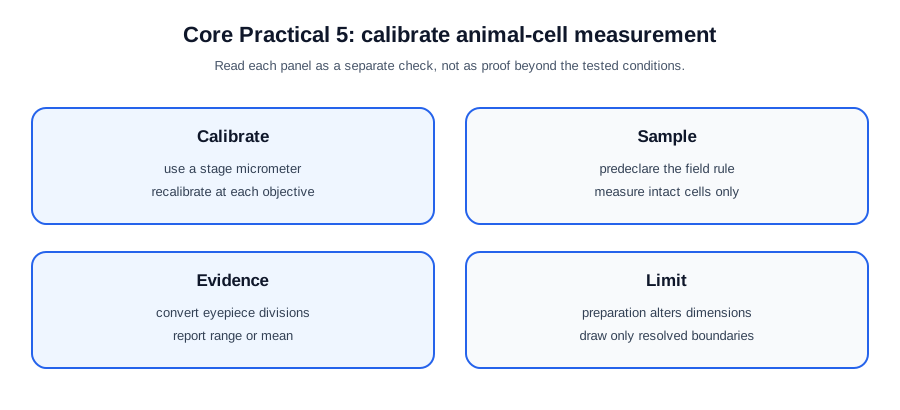
- Calibrate the eyepiece scale against a stage micrometer for each optical configuration used. Divide a known stage-micrometer length by the matching number of eyepiece divisions to obtain length per division. Apply that factor to cells selected by a stated field and eligibility rule, and record a clear drawing with a title and checked scale.
Check this topic from memory
Attempt the matching topic bank before reopening the notes. Use each missed idea to decide what to review next.
Start the topic quiz