

Pearson International A Level Biology Core Practical 6: Prepare and stain a root tip squash to observe mitosis
Pearson IAL Biology practical notes on prepare and stain a root tip squash to observe mitosis.
This guide practises root-tip preparation, observation and classification. It is not a complete validated staining or squashing protocol, or a claim to reproduce Pearson's prescribed procedure. Follow a named preparation method approved by your centre, with the current course documents and risk assessment; use a suitable prepared slide when preparation is not required.
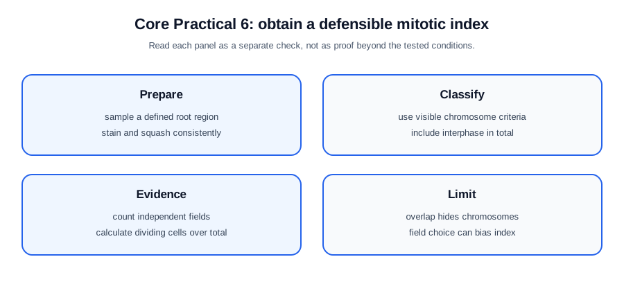
Aim and scientific principle
Observe chromosomes in an appropriate root-tip meristem preparation and identify cell-cycle stages using stated visible criteria. If required, estimate the mitotic index as a clearly defined fraction of the sampled cells; it is a snapshot fraction, not automatically a division rate or a stage duration.
State whether the task is descriptive observation, stage counting or a specified comparison. Descriptive observation and a mitotic-index estimate do not require a manipulated independent variable. For a comparison, define the treatment or tissue regions and the sampling unit before collecting data.
Apparatus and setup
Use a light microscope, the named root material, slides, coverslips and the tools and stain required by the approved preparation. Use an eyepiece graticule and stage micrometer if calibrated dimensions are required; counting stages alone does not require cell-length measurements. A ruler measures a drawing, not the microscopic specimen directly. Consider measuring range, resolution, uncertainty, calibration and suitability for the sample, not the instrument name alone. Draw a labelled scientific setup that shows the working geometry and exactly where readings are taken. Include a control or blank when it distinguishes the target effect from drift, contamination or background response.
Check focus, illumination and whether chromosomes can be distinguished under the chosen conditions. If measuring dimensions, calibrate the scale for the actual optical configuration and record reading uncertainty. A larger image or finer scale is not evidence of greater optical resolving power.
Method
- Select the species, root and defined meristem region using the approved protocol. Record root, slide and treatment identities. Follow its specified fixation or softening, staining and timing conditions rather than inventing an acid concentration or heating step.
- Where the protocol requires squashing, spread the tissue with controlled vertical pressure and avoid sliding the coverslip; use the prescribed protection against glass breakage. Alternatively, observe a suitable prepared slide when the task permits it.
- Locate the tissue at low power, centre it, then use appropriate higher power and fine focus without contacting the slide. Choose non-overlapping fields by a predeclared rule, with a boundary rule that prevents counting the same cell twice.
Check this topic from memory
Attempt the matching topic bank before reopening the notes. Use each missed idea to decide what to review next.
Start the topic quiz