

Pearson Edexcel International GCSE Human Biology Practical Skills 1: Microscopy and tissue observation
Pearson Edexcel International GCSE Human Biology Practical Skills 4HB1 notes on microscopy and tissue observation.
This chapter develops microscopy and observation skills supporting Pearson Edexcel International GCSE Human Biology (4HB1) Topic 1, especially recognition of cells, organelles and tissues in light and electron micrographs. It is an editorial practical-skills chapter, not a separately numbered Pearson practical. Theory owns cell and tissue functions; this hub owns observation, measurement, recording, safety and evaluation.
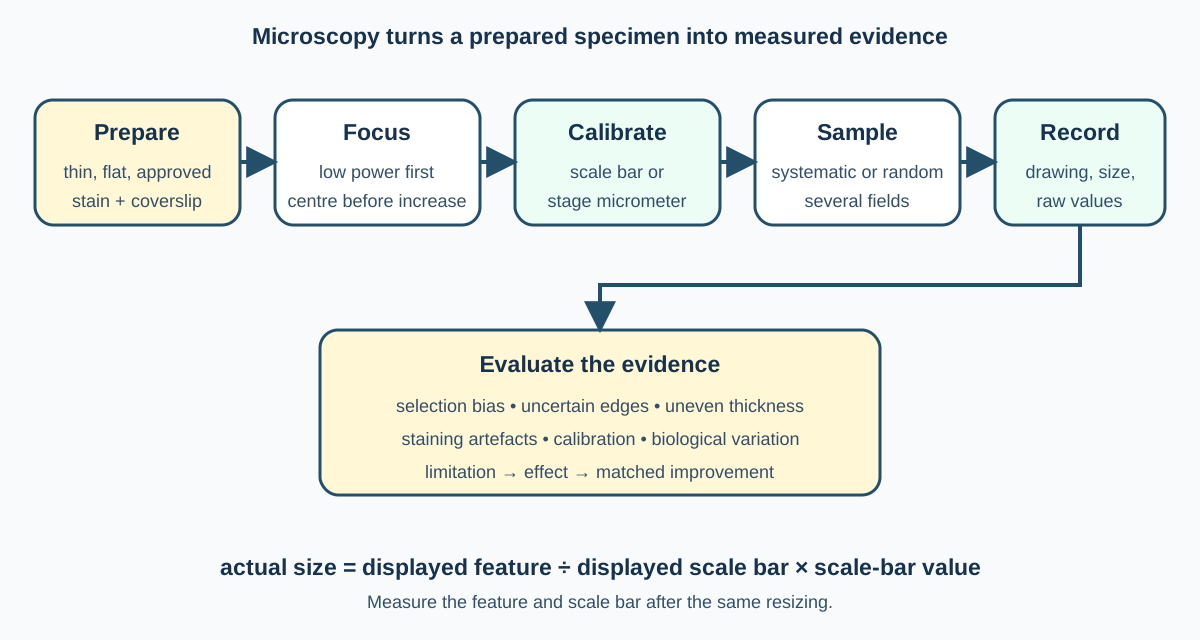
1. Turning a specimen into evidence
A useful specimen must be thin enough for transmitted light, flat enough to focus and prepared without avoidable distortion. Place a small approved specimen in a drop of water or suitable stain on a clean slide. Lower a coverslip at an angle with a mounted needle so air is pushed out rather than trapped.
Use only approved prepared material. Human blood, cheek cells or other body samples create consent, contamination and disposal issues and should not be collected unless an institution has an explicit supervised protocol. Prepared commercial slides provide safer repeatable material.
A stain increases contrast, not magnification. Different stains bind different structures, so colour in a prepared slide may be artificial evidence of treatment rather than natural tissue colour.
2. Focusing a light microscope
Begin with the lowest-power objective. Secure the slide and centre the specimen over the light. Watching from the side, bring the objective near the slide without contact. Then look through the eyepiece and use coarse focus to move away until the image appears. Adjust illumination and use fine focus for clarity.
Before changing to higher power, place the feature of interest in the centre. Higher power gives a smaller field of view, shallower depth of field and often less light. Use fine focus only at high power to reduce the risk of driving the lens into the slide.
Total magnification equals eyepiece magnification multiplied by objective magnification. Magnification makes an image larger; resolution determines whether two close points can be distinguished.
3. Biological drawings
A biological drawing records visible evidence rather than producing artwork. Use a sharp pencil, clear single lines and a large drawing. Do not shade. Show correct proportions and include only structures actually visible. Label with straight ruled lines that touch the structure and do not cross.
Add a title identifying specimen and view. Include magnification or, better, a scale bar. A drawing from one field is a selected observation and may not represent the whole tissue.
For tissue identification, use several features together. Epithelium forms sheets or linings; muscle contains elongated contractile cells; nervous tissue includes neurones and supporting cells; blood contains cells suspended in plasma. Shape alone is rarely conclusive without arrangement and context.
Check this topic from memory
Attempt the matching topic bank before reopening the notes. Use each missed idea to decide what to review next.
Start the topic quiz