

Pearson Edexcel International GCSE Human Biology Practical Skills 3: Diffusion and osmosis investigations
Pearson Edexcel International GCSE Human Biology Practical Skills 4HB1 notes on diffusion and osmosis investigations.
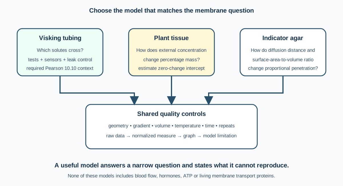
This chapter owns practical membrane evidence for Pearson Edexcel International GCSE Human Biology (4HB1), especially the required Visking-tubing investigation in outcome 10.10. Agar diffusion and tissue osmosis are supporting experimental models, not separately listed Pearson practicals. Theory owns diffusion, osmosis and active transport mechanisms; this page owns model choice, method, measurement, safety and evaluation.
1. What a membrane model can show
A partially permeable membrane lets some particles pass while restricting others by factors including molecular size and membrane properties. Visking tubing can model selective exchange, but it is not a living cell membrane. It has no transport proteins, ATP supply, blood flow or hormonal control.
An investigation should therefore ask a narrow question, such as whether glucose crosses while starch is retained, rather than claiming to reproduce a kidney or intestine completely.
2. Preparing Visking tubing
Soak the tubing as directed until flexible. Tie one end securely, add a measured solution without contaminating the outside, expel excess air and tie the second end. Rinse the outside thoroughly, blot consistently and check for leaks before immersion in a measured external solution.
Use equal tubing lengths and similar filled volumes in comparisons. Surface area, membrane thickness, concentration gradient, temperature, time and stirring affect exchange. A loosely tied knot can mimic diffusion by leaking bulk solution.
3. Starch and glucose model
Fill tubing with a starch-glucose mixture and immerse it in distilled water. Record initial tests on internal and external solutions. After a fixed time, retest both compartments.
Glucose may appear outside because its smaller particles cross the membrane. Starch should remain inside if the membrane excludes the larger molecules. Iodine added outside may diffuse inward and turn internal starch blue-black. The colour location and test sequence must be recorded carefully.
Use Benedict’s test with controlled heating for glucose and iodine for starch. Keep aliquots separate so one test reagent does not contaminate the other. Positive controls confirm reagents work; negative controls show the expected absence of reaction.
4. Quantifying diffusion
Qualitative colour change answers whether movement is detectable. Quantitative work can use a glucose sensor, colorimeter with calibration standards, mass change or timed sampling. Concentration should be inferred only from a validated calibration curve within its range.
Sample equal volumes at fixed times and replace removed external liquid if volume must remain constant, recording any dilution. A time-course graph can show fast initial change followed by a slower approach to equilibrium as the concentration gradient decreases.
Check this topic from memory
Attempt the matching topic bank before reopening the notes. Use each missed idea to decide what to review next.
Start the topic quiz