

Prepared microscope slides and plant sections: observation and handling

Understand a prepared slide, its label and coverslip, and distinguish visible tissue structure from live function.
A prepared slide carries a specimen in a form suitable for microscope observation. The glass slide supports it; a coverslip, where present, covers the preparation. Neither the glass outline nor a stain colour tells you which tissue you are observing.
This guide covers supplied preparations. Making fresh sections, adding stains and repairing mounts introduce different work and need their own supervised procedure.
Read the slide label before observation
Check the specimen name and the stated view before selecting a slide. A transverse section cuts across a structure; a surface preparation shows a different arrangement. Confirm that the slide matches the observation task rather than choosing by appearance alone.
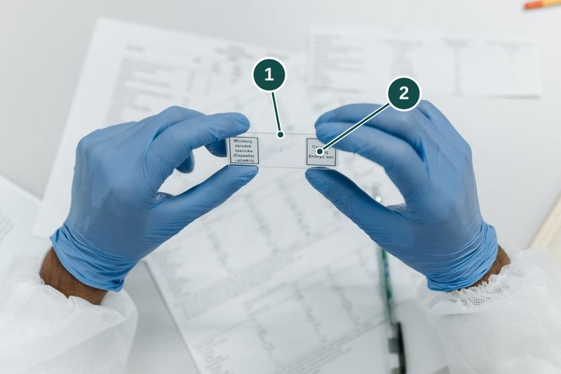
- Glass slide. Carries the preparation for observation.
- Specimen label. Names the preparation according to its label. Check that it matches the observation task.
Stock photograph for illustration by Pavel Danilyuk on Pexels. A prepared microscope slide with an identifying label. This example is labelled as a Capsella young embryo section. The photo does not show Eclat's specimens or demonstrate safe handling.
The photographed label names a Capsella young embryo section. Use this photo to recognise a slide and its label. It does not show a leaf or young stem section. We have not independently checked the specimen's species or preparation.