Cambridge International AS and A Level Physics 24: Medical physics
Cambridge International AS and A Level Physics 24: Medical physics
Study guide/
Cambridge Physics 9702 A Level notes on piezoelectric ultrasound, acoustic impedance, attenuation, X-ray production and contrast, CT reconstruction, positron annihilation and PE...
Medical physics is Topic 24 of the Cambridge International AS and A Level Physics 9702 additional A Level content. Sections 24.1 to 24.3 connect wave production, interaction and detection to ultrasound imaging, X-ray and CT contrast, and positron-emission tomography.
24.1 Production and use of ultrasound
Piezoelectric transduction
A piezoelectric crystal changes shape when a potential difference is applied across it. Conversely, changing its shape generates an e.m.f.
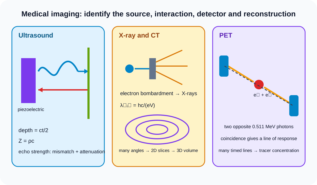
An alternating p.d. drives repeated expansion and contraction. If driven at a suitable high frequency, the crystal produces ultrasound waves. During detection, a returning pressure wave deforms the crystal and produces a varying e.m.f.
The same transducer can emit a short pulse and then switch to receiving echoes. A backing material can limit prolonged vibration, but detailed transducer construction is not required here.
Ultrasound has frequency above the upper limit of human hearing. Its diagnostic value comes from pulse timing, reflection and attenuation, not merely from being inaudible.
Pulse-echo imaging
An emitted pulse travels through tissue and partially reflects at boundaries where acoustic properties change. The transducer detects echoes after a time delay.
For a boundary at depth d and wave speed c in the intervening medium:
2d=ct,
so
d=2ct.
Check this topic from memory
Attempt the matching topic bank before reopening the notes. Use each missed idea to decide what to review next.
The factor two accounts for outward and return travel. If speed varies along the route, a single-speed estimate becomes an approximation.
Echo delay gives boundary position; echo magnitude gives information about boundary contrast and attenuation. Repeated pulses along different directions build an image of internal structures.
Acoustic impedance and reflection
Specific acoustic impedance Z of a medium is
Z=ρc,
where rho is density and c is sound speed in the medium. Its unit is kilogram per square metre per second.
For normal incidence at a boundary between impedances Z1 and Z2, intensity reflection coefficient is
I0IR=(Z1+Z2Z1−Z2)2.
The ratio is a fraction between zero and one in the ideal model. Equal impedances give zero reflected intensity. A greater mismatch gives stronger reflection.
The order of Z1 and Z2 does not change the squared coefficient. The reflected wave phase can depend on direction of mismatch, but the official equation concerns intensity magnitude.
Coupling gel reduces the large impedance mismatch that an air gap would create between transducer and skin, allowing more ultrasound energy to enter the body.
Attenuation
Ultrasound intensity decreases with distance x through matter:
I=I0e−μx,
where mu is the attenuation coefficient. Its reciprocal-length unit must match x.
Attenuation includes energy removal from the original beam by absorption and scattering. Greater mu or greater path length gives smaller transmitted intensity.
For a round trip to depth d in one medium, total path is approximately 2d. Do not insert one-way depth into the attenuation exponent if the question asks for the returning echo after both journeys.
Taking logarithms gives a straight-line form:
ln(I/I0)=−μx.
Reflection and attenuation are separate effects. First decide how much reaches a boundary, then apply reflection, then attenuate the returning portion if the complete echo intensity is required.
24.2 Production and use of X-rays
X-ray production
Electrons are accelerated through a high potential difference and bombard a metal target. Their rapid energy changes produce X-rays while much input energy becomes thermal energy in the target.
An electron accelerated through p.d. V gains maximum kinetic energy eV. If all that energy becomes one photon:
eV=hfmax=λminhc.
Therefore,
λmin=eVhc.
This is the minimum wavelength, corresponding to maximum photon energy. Most electrons do not convert all kinetic energy into one photon, so the spectrum contains longer wavelengths too.
Increasing accelerating p.d. decreases minimum wavelength. It does not mean every emitted photon has that wavelength.
Contrast and attenuation
X-rays pass through the body and are attenuated by different amounts in different tissues. A detector records transmitted intensity.
For homogeneous material:
I=I0e−μx.
Greater thickness or attenuation coefficient gives less transmitted intensity.
Contrast is a difference in recorded intensity or image brightness between structures. It arises when tissues have different attenuation or path thickness. Strong contrast makes boundaries or structures easier to distinguish.
For several tissue layers, attenuation factors multiply, equivalent to adding the products mu x in the exponent:
I=I0e−(μ1x1+μ2x2+⋯).
A conventional projection image superposes all structures along each ray path. This can hide depth information.
CT reconstruction
Computed tomography first combines multiple X-ray projections of the same section taken from different angles to reconstruct a two-dimensional image of that section.
The process is repeated for sections along an axis. The set of two-dimensional slice images is then combined into a three-dimensional representation.
Do not describe CT as one ordinary X-ray photograph rendered in 3D. The angular projection data and repeated slices are essential.
Each reconstructed pixel or voxel represents information related to local attenuation. CT improves separation of overlapping structures compared with a single projection.
Detailed reconstruction algorithms and clinical protocol are outside the syllabus boundary.
24.3 PET scanning
Tracer and beta-plus decay
A tracer is a substance containing radioactive nuclei introduced into the body and absorbed by the tissue being studied. Its distribution follows the biological process associated with the tracer.
PET uses a tracer whose nuclei undergo beta-plus decay and emit positrons. The tracer should allow useful detection while limiting unnecessary persistence, but isotope-selection detail is beyond the stated outcomes.
Annihilation
A positron is the electron's antiparticle. After losing kinetic energy in tissue, it interacts with an electron. They annihilate, converting mass-energy into photons while conserving total energy and momentum.
If the electron-positron pair is approximately at rest before annihilation, momentum conservation requires two gamma photons in opposite directions. One photon alone could not conserve zero total momentum.
Total rest energy is
Etotal=2mec2.
The two photons share it equally under the at-rest approximation:
Eγ=mec2.
Each photon has about 0.511 MeV. If the pair has residual momentum, the exact laboratory energies and directions can differ slightly, but the syllabus model uses the opposite equal-photon case.
Coincidence detection and image formation
The gamma photons leave the body and can be detected in a surrounding detector ring. Detectors on approximately opposite sides recording photons within a short timing interval identify a coincidence event.
The annihilation is inferred to lie along the line connecting the detector pair. A single event does not locate one exact point on that line.
Processing many coincidence lines and their arrival times reconstructs the spatial concentration of tracer in tissue. Regions with more tracer activity contribute more events.
PET maps tracer concentration and therefore functional or metabolic information associated with the tracer; it is not simply an anatomical photograph made directly by the positron.
Random or scattered coincidences can reduce image quality in real systems, but detailed corrections are beyond this boundary.
Comparing the modalities
Ultrasound uses mechanical waves and echoes from impedance boundaries. X-ray and CT use differential attenuation of electromagnetic radiation. PET detects paired annihilation photons from radioactive tracer distribution.
Choose equations by modality. The exponential attenuation form occurs for both ultrasound and X-rays, but their wave types, sources, interaction details and imaging signals differ.
Theory and practical ownership
This theory note covers the transduction, reflection, attenuation, photon-energy and reconstruction principles specified by Topic 24.
Practical or clinical execution covers transducer coupling, beam alignment, detector calibration, exposure optimisation, tracer handling, acquisition timing, uncertainty and safety. No clinical recommendation is being made; these are examination-physics models.
Worked application: ultrasound echo and PET photon energy
An ultrasound pulse travels through tissue at 1540m⋅s−1 and returns after 130us, placing the boundary at ct/2=0.100m. If outward and return attenuation coefficient is 8.0m−1, the path factor is e−8.0(0.200)=0.202 before applying boundary reflection. In PET, an approximately stationary electron and positron have combined rest energy 2mec2=1.022MeV. Momentum conservation gives two opposite photons, each with 0.511MeV. Echo timing locates a boundary, while coincidence lines localise tracer events statistically.
Common misconceptions and corrections
Saying a piezoelectric crystal only emits ultrasound. It also generates e.m.f. when deformed.
Applying constant p.d. and expecting a sustained wave. Alternating deformation generates ultrasound.
Calling ultrasound electromagnetic radiation. It is a mechanical wave.
Using ct as echo depth. Divide the round-trip distance by two.
Ignoring changes in sound speed between tissues. The simple depth estimate assumes a route speed.
Using density alone as acoustic impedance. Z equals rho c.
Calling impedance a resistance measured in ohms. Its acoustic unit differs.
Saying equal impedances give maximum reflection. They give zero in the ideal coefficient.
Forgetting to square the impedance ratio. The equation is for intensity.
Saying coupling gel increases an air gap. It reduces impedance mismatch.
Using one-way distance for a returning echo's attenuation. Include both path legs.
Treating reflection and attenuation as the same calculation. Apply their factors separately.
Using a positive attenuation exponent. Intensity decays with distance.
Saying X-rays arise from the target merely being hot. Accelerated electrons bombard it.
Equating eV to every X-ray photon energy. It is the maximum available per electron.
Calling hc/eV the maximum wavelength. It gives the minimum.
Saying higher accelerating voltage increases minimum wavelength. It decreases it.
Calling contrast total image brightness. It is difference between structures.
Saying greater attenuation gives greater transmitted intensity. It gives less.
Adding intensities through layers instead of exponents. Attenuation factors multiply.
Describing CT as one projection. It combines many angles for each slice.
Saying one reconstructed slice is already the complete 3D image. Multiple slices are combined.
Calling PET tracer a detector. It is a radioactive substance absorbed by studied tissue.
Using beta-minus tracer decay for PET. The stated process is beta-plus.
Saying a positron is a proton. It is the electron antiparticle.
Saying annihilation destroys energy. Mass-energy and momentum are conserved.
Producing one photon from an at-rest electron-positron pair. It cannot conserve zero momentum.
Giving the two photons parallel directions. They travel oppositely in the ideal case.
Assigning 1.022 MeV to each photon. That is the pair total; each is about 0.511 MeV.
Saying one coincidence fixes an exact annihilation point. It defines a line of response.
Saying PET directly photographs positrons. It detects external gamma photons.
Calling PET only anatomical imaging. It maps tracer concentration and associated function.
Using ultrasound impedance reflection for X-ray contrast. The modalities use different interactions.
Treating examination models as clinical advice. They explain physical principles only.
Assessment guidance
Identify the modality before selecting its signal and equation. For ultrasound, trace generation, outward propagation, boundary reflection, return attenuation and piezoelectric detection, using total echo path. For X-rays, equate electron energy eV to maximum photon energy only for minimum wavelength, and explain contrast through differential attenuation. Describe CT in the required order: multiple angles to one 2D slice, repeated slices, then 3D combination. For PET, conserve mass-energy and momentum to obtain two opposite photons, then distinguish a coincidence line from an exact point and many processed events from one detection.
Retrieval practice
Derive echo depth, calculate impedance reflection and combine outward and return attenuation. Calculate X-ray minimum wavelength and compare transmitted intensities through layered tissues. Explain CT reconstruction without using the phrase 3D X-ray alone. Follow a PET event from tracer uptake through beta-plus decay, annihilation, paired detection, coincidence line and tracer-concentration image.