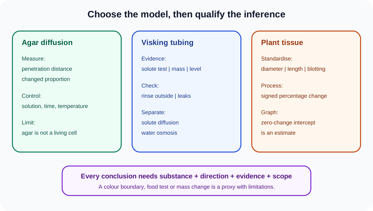
Diffusion and osmosis are explicit Cambridge IGCSE Biology practical contexts for Papers 5 and 6. Strong investigation answers identify the moving substance, the measured proxy, the controlled gradient and the limits of each model rather than treating every membrane result as osmosis.
Choose a model that answers the question
Diffusion can be investigated through agar, liquid or a partially permeable membrane. Osmosis requires water moving through a partially permeable membrane from higher water potential to lower water potential.
Plant tissue provides living cell membranes and a measurable mass or length change. Visking tubing provides a non-living partially permeable barrier. Agar can reveal diffusion distance or proportion reached by a substance but does not model every feature of a cell.
State what the apparatus represents and what it does not. A model supports a specific inference, not proof that a living organism behaves identically.
Diffusion through agar
A coloured or indicator-containing agar block can be placed in a solution that changes its colour as diffusion occurs. Measure block dimensions before exposure, keep solution concentration, volume, temperature and time constant, then cut the block to measure penetration.
To compare block size, use cubes made from the same agar batch and expose them under identical conditions. Smaller cubes have a larger surface-area-to-volume ratio and may have a greater proportion reached in the same time.
Diffusion distance can be measured from each face, or the undiffused core can be measured. Define the rule before collecting data. A percentage or volume proportion can compare different starting sizes more fairly than raw penetration distance alone.
Use forceps and a cutting tile. If acid, alkali or indicator is involved, follow the supplied hazard information and wear eye protection.
Visking-tubing investigations
Soak and open the tubing as instructed, secure one end, add the selected solution and tie the other end. Rinse the outside thoroughly to remove material spilled during filling, check for leaks and place the tubing in a known external solution.
After a controlled time, test the liquid inside or outside using the appropriate reagent. A positive test outside supports that the tested molecule crossed the membrane. A negative result may mean the molecule did not cross, was below detection level or the method was insufficiently sensitive.
Keep tubing length or surface area, internal and external volumes, concentration, temperature and time constant when comparing conditions.
Small molecules may pass while larger molecules do not, depending on membrane pores. Do not state that a molecule is too large without evidence from the supplied results.
Check this topic from memory
Attempt the matching topic bank before reopening the notes. Use each missed idea to decide what to review next.
Mass or liquid-level change may provide evidence of net water movement, while a food test provides evidence about a solute. Separate these observations and mechanisms.
Prevent false transfer results
Failure to rinse the outside after filling can produce a false positive in the surrounding solution. A leak can allow bulk mixture to escape rather than selective movement across the membrane.
Use a negative control with no target substance outside and, where useful, a positive control to confirm the food-test reagent works. Check knots and handle the tubing consistently.
Sampling the surrounding liquid before and after the investigation can distinguish initial contamination from later transfer.
Plant-tissue osmosis method
Prepare at least five solution concentrations across a useful range. Cut potato or other plant cylinders with the same cork borer and to the same measured length. Remove skin where instructed so surfaces are comparable.
Record initial mass using the same balance. Place each cylinder in an equal volume of solution for the same time at the same temperature. Ensure tissue is immersed.
Remove cylinders, blot each using the same material, pressure and duration, then measure final mass promptly. Inconsistent surface liquid can overwhelm the biological mass change.
Use several independent cylinders per concentration. They are biological replicates, not merely repeated balance readings of one cylinder.
Calculate percentage change
Use:
percentage change = (final mass - initial mass) / initial mass × 100
A positive result shows mass gain; a negative result shows mass loss. Percentage change allows specimens with different initial masses to be compared.
Do not discard the sign. Absolute change loses direction and can make water entry and water loss look identical.
Record raw initial and final masses, calculated percentage changes, replicate values and a mean. Round consistently and retain enough precision for the trend.
Estimate the zero-change concentration
Plot solution concentration on the x-axis and mean percentage mass change on the y-axis. Draw an appropriate best-fit line or curve.
The x-intercept, where percentage change is zero, estimates the external concentration producing no net mass change under those conditions. Interpolate between points when needed.
This is an estimate, not an exact universal value for every potato. Variety, tissue location, age, temperature and method affect results. Use closer concentration intervals around the intercept to refine the estimate.
No net mass change does not mean water molecules stop moving. Movement continues in both directions at equal overall rates.
Investigate factors affecting diffusion
Temperature, concentration gradient, diffusion distance and surface-area-to-volume ratio can be varied one at a time. Define a measurable dependent variable such as time to a colour endpoint, penetration distance in a fixed time or proportion of a block changed.
When temperature is varied, equilibrate solutions and apparatus before starting and monitor actual temperature. When concentration is varied, keep total volume and specimen dimensions constant.
Do not change block size and surface-area-to-volume ratio as if they were independent unrelated variables. Size causes the ratio change.
Interpret results using the correct process
Net water entry into plant cells occurs when external water potential is higher than cell water potential. Cells become turgid and tissue may gain mass. In a more concentrated external solution, water leaves cells, tissue loses mass and cells may become flaccid.
Solute movement down its concentration gradient is diffusion, not osmosis. Water movement through the membrane is osmosis.
In agar, colour change may indicate reaction after a diffusing substance arrives. The measured colour boundary is a proxy for diffusion, not the movement itself.
Evaluate the investigation
Unequal tissue size changes surface area, volume and initial mass. Use one cork borer and a cutting guide. Tissue from different potato regions may vary; randomise or distribute pieces across conditions and use replicates.
Inconsistent blotting changes surface liquid; standardise the method. Evaporation changes solution concentration; cover vessels. Temperature drift changes particle movement; use a controlled environment.
Wide concentration intervals weaken the intercept estimate; test narrower intervals near zero change. A balance with coarse resolution may not detect small changes; use suitable precision.
More repeats improve reliability but do not correct a leak, contaminated tubing or confounded variables.
Worked application: locate an osmosis intercept
Potato cylinders in 0.20 mol/dm³ solution show a mean mass change of +5.0%, while cylinders in 0.30 mol/dm³ show -3.0%. The zero-change concentration lies between these values. A graph-based interpolation gives an estimate near 0.26 mol/dm³, depending on the fitted relationship. This does not prove that potato cells contain a 0.26 mol/dm³ solution, because cell contents are mixtures and the result depends on tissue and method. Repeat with 0.24, 0.26 and 0.28 mol/dm³, standardised cylinders and consistent blotting to refine the estimate.
Common misconceptions and corrections
Calling every movement across tubing osmosis. Solutes diffuse; water moves by osmosis.
Saying osmosis is movement of solution. It is net water movement through a partially permeable membrane.
Saying particles stop at equilibrium. Random movement continues.
Treating Visking tubing as a living cell. It is a membrane model.
Assuming every small molecule crosses. Use evidence from the test.
Calling a negative food test proof of no crossing. Concentration may be below detection.
Forgetting to rinse filled tubing. External contamination can cause a false positive.
Ignoring leaks. Bulk escape is not selective transfer.
Using different tubing areas. Transfer opportunity changes.
Calling agar colour change osmosis. It commonly tracks diffusion and reaction.
Comparing different agar sizes using only raw changed volume. Use a fair proportion where needed.
Changing block size and solution concentration together. Isolate one variable.
Cutting potato cylinders with different diameters. Use one cork borer.
Using equal lengths but leaving skin on some pieces. Standardise exposed surfaces.
Using different solution volumes. Concentration may change differently.
Leaving some tissue partly exposed. Ensure comparable immersion.
Blotting some cylinders more strongly. Standardise surface-liquid removal.
Using final mass alone. Initial mass is required.
Comparing raw mass change for unequal starting masses. Use percentage change.
Removing the negative sign. Direction is biologically meaningful.
Saying positive change means solute entered. It normally indicates net water entry.
Saying zero change means no water movement. There is no net change.
Calling the intercept exact. It is an estimate under tested conditions.
Using only two concentrations to define a curve. Test several values.
Claiming the highest tested concentration is an optimum. Osmosis here has no rate optimum claim.
Using repeats to fix contaminated tubing. Correct the method.
Generalising one potato to all plant tissue. Biological variation limits scope.
Describing water movement without a relative potential. State the direction and membrane.
Assessment guidance
Name the moving substance and measured proxy before explaining a result. Visking-tubing answers should include leak checks, external rinsing, controlled area, volumes, concentration, time and an appropriate test. Plant-tissue plans need standard dimensions, initial and final measurements, consistent blotting, equal solution conditions and biological replicates. Preserve percentage-change signs and use a graph intercept only as an estimate of zero net change. Evaluation marks require limitation-effect-improvement chains, such as unequal cylinders changing exchange area and a cork borer plus cutting guide correcting it.
Retrieval practice
Design one agar, one Visking-tubing and one potato investigation, naming the moving substance, dependent measure and four controls. Calculate signed percentage changes and estimate an x-intercept from a graph. Diagnose false positives, leaks, inconsistent blotting, evaporation and wide concentration intervals, then write a targeted improvement for each without confusing diffusion with osmosis.
Theory and practical ownership
This practical note owns diffusion and osmosis models, apparatus, variables, calculations, graphs, controls, safety and evaluation. The movement-in-and-out-of-cells theory note owns definitions and cell-level mechanisms. The data-skills note owns general graph construction.