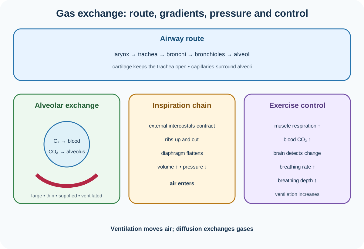
Topic 11 of Cambridge IGCSE Biology 0610 and 0970 separates ventilation, which moves air, from gas exchange, which moves oxygen and carbon dioxide by diffusion. The official section covers exchange-surface features, breathing-system anatomy, inspired and expired air, exercise responses and airway protection. Smoking is not imported into this Topic 11 boundary.
The breathing-system route
Air passes through the larynx into the trachea. The trachea divides into two bronchi, one entering each lung. Bronchi branch into narrower bronchioles, which lead to alveoli. Alveoli are closely associated with blood capillaries.
Rings of cartilage in the trachea support the airway and prevent it collapsing during pressure changes while leaving the passage open for airflow. The syllabus names cartilage function in the trachea, so do not generalise it as the structure that performs gas exchange.
The lungs occupy the thorax and are ventilated through movements of the ribs, intercostal muscles and diaphragm. The lungs themselves do not contain muscles that actively pull them open in the required explanation.
Gas-exchange surface features
Alveoli collectively provide a large surface area. A large area permits many oxygen and carbon dioxide molecules to diffuse at the same time.
The alveolar surface and adjacent capillary wall are thin, creating a short diffusion distance. A good blood supply continually brings deoxygenated blood and carries oxygenated blood away. Good ventilation renews alveolar air.
Together, blood flow and ventilation maintain concentration gradients. Oxygen concentration is higher in alveolar air than in deoxygenated blood, so oxygen diffuses into blood. Carbon dioxide concentration is higher in blood arriving at the lungs than in alveolar air, so carbon dioxide diffuses into alveoli.
Gas exchange is diffusion down concentration gradients. Ventilation maintains those gradients but is not itself diffusion across the exchange surface.
Inspiration
During inspiration, external intercostal muscles contract. The ribs move upward and outward. The diaphragm muscles contract, making the diaphragm flatten and move downward.
These actions increase thoracic volume. As volume increases, pressure inside the thorax and lungs falls below atmospheric pressure. Air moves into the lungs down the pressure difference.
Use the complete causal order:
muscle contraction→rib and diaphragm movement→volume increase→pressure decrease→air enters.
Check this topic from memory
Attempt the matching topic bank before reopening the notes. Use each missed idea to decide what to review next.
The external intercostal muscles do not push air directly. They change rib position, which changes volume and pressure.
Expiration
During normal expiration, external intercostal muscles relax, the ribs move downward and inward, and the diaphragm relaxes and becomes more dome-shaped. Thoracic volume decreases, pressure rises above atmospheric pressure and air moves out.
The Supplement includes internal and external intercostal muscle identification. Internal intercostal muscles can contract during forced expiration, pulling ribs downward and inward more strongly. For ordinary quiet expiration, elastic recoil and relaxation are central.
Air moves because of a pressure difference. Saying simply that "the diaphragm pushes air out" omits the volume-pressure mechanism.
Inspired and expired air
Inspired air contains more oxygen than expired air because oxygen diffuses into blood and is used in aerobic respiration. Expired air still contains oxygen; not all inhaled oxygen is absorbed.
Expired air contains more carbon dioxide because respiring cells produce carbon dioxide, blood transports it to lungs and it diffuses into alveoli.
Expired air contains more water vapour because air passes over moist breathing surfaces and gains water. Nitrogen proportion changes little and is not one of the three required comparison substances in this section.
Limewater tests for carbon dioxide. Air with more carbon dioxide turns limewater cloudy more quickly under comparable conditions. In a valid comparison, equal air volumes should pass through equal limewater volumes at similar flow rates. Detailed assembly and hygiene belong to the practical hub.
Explain the composition differences
Oxygen is removed from alveolar air by diffusion into capillary blood. It binds to haemoglobin and is transported to tissues for aerobic respiration.
Carbon dioxide is produced by respiration in cells, transported in blood and diffuses from blood into alveoli. Ventilation then removes it during expiration.
Water evaporates from moist airway and alveolar surfaces, increasing the water-vapour content of expired air. Temperature and surrounding humidity can affect measured water condensation, so interpret the observation carefully.
Physical activity and breathing
During physical activity, muscle contraction requires more energy. Aerobic respiration increases and produces carbon dioxide faster. Carbon dioxide concentration in blood rises.
The brain detects the increased carbon dioxide concentration and causes breathing to become faster and deeper. Increased rate moves breaths more frequently; increased depth increases air volume per breath. Together they raise ventilation, bring in more oxygen and remove carbon dioxide faster.
The required control chain is:
activity→respiration increases→blood carbon dioxide rises→brain detects change→rate and depth increase.
Do not explain the response only by saying that muscles "need more air". The syllabus specifically requires increased carbon dioxide in blood, detection by the brain and the resulting rate and depth change.
Goblet cells, mucus and ciliated cells
Goblet cells secrete mucus. Mucus traps pathogens and particles before they reach delicate gas-exchange surfaces.
Ciliated cells have cilia that beat and move the mucus toward the throat, where it can be swallowed. Stomach acid then destroys many swallowed microorganisms.
The components form a sequence: goblet cell produces mucus, mucus traps and cilia move the trapped material. Cilia do not produce mucus and mucus does not actively beat.
This protection maintains clear airways and reduces pathogen and particle entry. It is distinct from alveolar gas exchange itself.
Interpret breathing evidence
Breathing rate is breaths per unit time. Breathing depth is volume moved in one breath. Ventilation per minute depends on both:
minute ventilation=breathing rate×volume per breath.
A person taking 12 breaths per minute at 0.5 dm³ per breath ventilates 6.0 dm³ per minute. If rate rises but depth falls, total ventilation cannot be inferred without both values.
Exercise investigations require standardised activity, defined counting intervals, safe participant screening, recovery and repeats. They estimate a response but are influenced by fitness, effort, temperature and measurement timing.
Worked application: explain ventilation and exercise
Before exercise, a student breathes 12 times per minute with an estimated depth of 0.50 dm³, giving 6.0 dm³ per minute. After a standardised activity, rate is 24 per minute and depth is 0.80 dm³, giving 19.2 dm³ per minute. Working muscles respire faster and increase blood carbon dioxide concentration. The brain detects this increase and raises breathing rate and depth, increasing oxygen intake and carbon dioxide removal. During each inspiration, external intercostal and diaphragm muscles contract, thoracic volume increases, pressure falls below atmospheric pressure and air enters. The calculation therefore supports increased ventilation, not faster diffusion caused directly by muscle movement.
Common misconceptions and corrections
Calling ventilation gas exchange. Ventilation moves air; gas exchange is diffusion across a surface.
Starting the airway route at alveoli. Air travels through larynx, trachea, bronchi and bronchioles first.
Calling bronchioles the two main branches of the trachea. Those are bronchi.
Saying cartilage carries oxygen. It supports the trachea and keeps it open.
Saying lungs contain the muscles that pull air in. Intercostal and diaphragm muscles change thoracic volume.
Calling an alveolus a blood vessel. It is an air sac associated with capillaries.
Saying alveoli have a small surface area. Their large collective area increases exchange.
Saying expired air contains no oxygen. It contains less oxygen than inspired air.
Saying inspired air contains no carbon dioxide. It contains much less, not none.
Saying expired air contains less water vapour. It generally contains more.
Calling cloudy limewater an oxygen result. It indicates carbon dioxide.
Comparing limewater without controlling air volume. Unequal volumes weaken the inference.
Saying respiration occurs in lungs only. Cells throughout the body respire.
Explaining exercise breathing only through oxygen demand. Include blood carbon dioxide and brain detection.
Calling breathing rate the volume of each breath. That is depth.
Saying deeper breathing means more breaths per minute. Depth and rate are separate.
Adding rate and depth to calculate ventilation. Multiply them.
Saying goblet cells beat mucus upward. Ciliated cells move it.
Saying ciliated cells secrete mucus. Goblet cells secrete it.
Saying mucus actively kills every pathogen. Its named role is trapping.
Importing smoking outcomes into Topic 11. They are not in the official section 11.1 boundary.
Assessment guidance
Separate anatomy, ventilation and gas exchange. Trace air in order, then pair each alveolar feature with surface area, diffusion distance or gradient maintenance. For breathing mechanics, always write muscle action, rib or diaphragm movement, thoracic volume, pressure and airflow in that order. Compare inspired and expired air using oxygen, carbon dioxide and water vapour, then explain each difference through respiration, blood transport or moist surfaces. In exercise answers, include increased blood carbon dioxide, brain detection and increased rate and depth. For airway defence, assign secretion to goblet cells, trapping to mucus and movement to ciliated cells.
Retrieval practice
Draw and label the complete breathing route and an alveolus-capillary interface. Build matched inspiration and expiration chains from muscle to airflow, compare the three required air components and calculate minute ventilation from four rate-depth pairs. Finish by reconstructing the physical-activity control loop and goblet-mucus-cilia protection sequence.
Theory and practical ownership
This theory note owns breathing anatomy, exchange-surface adaptations, pressure-driven ventilation, air-composition explanations, exercise control and airway protection. The separate Biology practical hub owns limewater apparatus, equal-volume comparison, breathing-rate and depth measurement, exercise standardisation, participant safety, recovery, repeats, tables, graphs and evaluation.