

Pearson Edexcel International GCSE Human Biology 4: Bones, muscles and joints

Study guide

Pearson Edexcel International GCSE Human Biology 4HB1 notes on bones, muscles and joints.

Movement depends on a living support system, not on bones or muscles acting alone. Pearson Edexcel International GCSE Human Biology (4HB1) Topic 4 covers the axial and appendicular skeletons, long-bone organization, elbow, shoulder and intervertebral joints, synovial-joint structure, antagonistic action of biceps and triceps, dietary support for bone and muscle, and osteoporosis. The topic links anatomy to force transmission, stability, repair and disease risk.
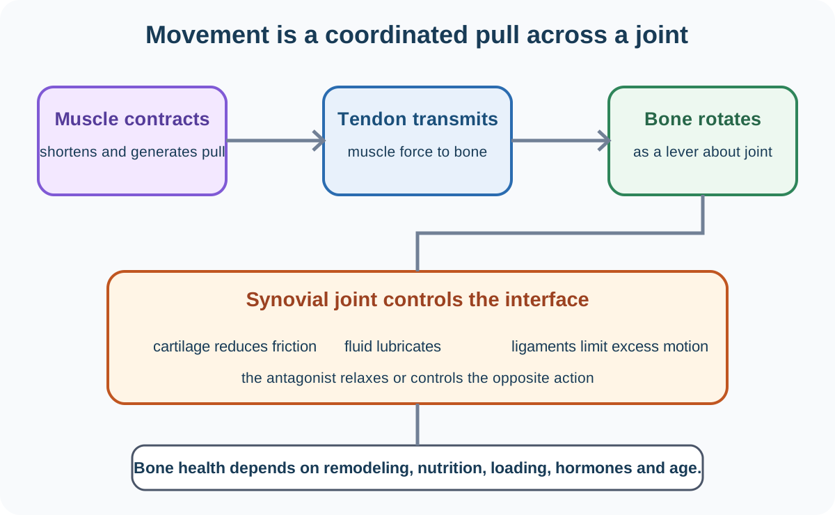
A musculoskeletal movement map connecting skeleton, synovial joints and antagonistic muscles

1. Functions of the skeleton

The skeleton supports soft tissues, protects organs, provides attachment and levers for muscles, stores minerals and contains marrow involved in blood-cell production. It is living tissue that remodels in response to load and physiological signals.

The axial skeleton forms the central axis: skull, vertebral column and ribcage. The skull protects the brain; vertebrae protect the spinal cord while allowing controlled movement; ribs and sternum protect thoracic organs and contribute to breathing mechanics.

The appendicular skeleton includes shoulder girdle, pelvic girdle and limbs. The scapula and clavicle position the upper limb and allow wide movement. The pelvis transfers body weight to lower limbs and protects pelvic organs. Limb bones act as levers around joints.

Axial does not mean “all bones near the middle”, and appendicular does not mean only hands and feet. Use the named components required by Pearson.

2. Long-bone structure

A long bone such as the femur has a shaft and expanded ends called epiphyses. Compact bone forms a strong dense outer region, especially around the shaft, resisting bending and compression. Spongy bone forms an internal lattice, prominent toward the epiphyses, providing strength with lower mass and containing marrow spaces.

Articular cartilage covers joint surfaces at epiphyses, reducing friction and absorbing shock. The central shaft contains a marrow cavity. Blood vessels and living cells support growth, repair and remodeling.

Bone architecture distributes material rather than making the whole bone solid. A completely solid bone would be heavy, while a thin hollow shell without internal organization would resist some loads poorly.

3. Joint types and examples

A joint is where two or more bones meet. Its structure balances movement and stability.

The elbow is mainly a hinge synovial joint, allowing flexion and extension in one principal plane. The shoulder is a ball-and-socket synovial joint, allowing movement in many directions and rotation. Its mobility comes with less inherent stability than the deeper hip joint.

Intervertebral joints include cartilaginous connections between vertebral bodies. Intervertebral discs allow limited movement, absorb shock and distribute compression. Small movements across many vertebrae combine to produce flexibility while protecting the spinal cord.

Do not call every joint synovial or assume all synovial joints move equally. Shape, ligaments and surrounding muscles constrain range.

4. Synovial-joint structure

In a synovial joint, articular cartilage covers bone ends and reduces friction. A joint capsule surrounds the joint. Its fibrous outer layer helps hold bones together, while an inner synovial membrane produces synovial fluid.

Synovial fluid lubricates surfaces and helps nourish avascular cartilage. Ligaments connect bone to bone and limit excessive movement. Tendons connect muscle to bone and transmit muscle force. These structures have different roles and should not be interchanged.

The joint cavity permits low-friction movement. Damage to cartilage or altered fluid and inflammation can make movement painful and reduce function.

5. Muscles, tendons and levers

Voluntary skeletal muscles attach to bones through tendons. Muscle contraction generates pulling force; muscles cannot actively push bones. Therefore joints are controlled by antagonistic pairs whose actions oppose one another.

At the elbow, biceps contraction and triceps relaxation flex the forearm. The biceps tendon pulls on the radius. For extension, triceps contracts while biceps relaxes, and the triceps tendon pulls on the ulna.

The elbow acts as a pivot, bones as levers and muscle force as effort. Because muscle attachment may lie close to the joint, a large muscle force can produce a smaller but faster movement at the hand over a greater distance.

Stabilizing muscles around the shoulder keep the humeral head aligned while larger muscles move the arm. Movement is coordinated rather than a simple single-muscle switch.

6. Dietary support for muscle and bone

Protein provides amino acids for muscle proteins, enzymes and tissue repair. Adequate energy intake prevents excessive use of body protein as fuel. Calcium and phosphate contribute to bone mineral, while vitamin D supports calcium absorption and normal mineralization.

Vitamin C supports collagen formation, important in connective tissues and bone matrix. A balanced diet also supplies other micronutrients. Nutrition acts with mechanical loading, hormones, age and health; taking one nutrient alone does not guarantee strong bones or large muscles.

Weight-bearing activity stimulates bone maintenance and supports muscle strength. Prolonged inactivity reduces loading signals and can accelerate loss.

7. Bone growth and remodeling

During growth, cartilage regions near long-bone ends contribute to lengthening before maturity. Bone-forming and bone-resorbing cells continually remodel adult bone, repairing microdamage and adapting structure.

Bone mass usually rises during growth toward a peak, then maintenance depends on the balance between formation and resorption. Hormones, nutrition, exercise and disease influence this balance.

A fracture heals through coordinated clotting, tissue formation, new bone deposition and remodeling. Healing requires blood supply and appropriate stabilization.

8. Osteoporosis

Osteoporosis is a condition in which bone mass and microarchitecture deteriorate, making fractures more likely. It may progress without obvious symptoms until a low-impact fracture occurs.

Risk factors include increasing age, reduced estrogen after menopause, family history, low body mass, inactivity, smoking, excessive alcohol, low calcium or vitamin D status and some medicines or diseases. A risk factor changes probability; it does not mean every exposed person develops the condition.

Possible effects include fractures of hip, wrist or vertebrae, back pain, loss of height and altered posture. Vertebral compression can occur without a dramatic injury.

Prevention and management can include weight-bearing and resistance exercise, adequate calcium and vitamin D, avoiding smoking and excessive alcohol, fall-risk reduction and prescribed medication. Advice must be individualized clinically rather than presented as a guaranteed cure.

9. Integrating structure and movement

For any movement explanation, trace the chain:

  1. a named muscle contracts and shortens;
  2. its tendon transmits pull to a named bone;
  3. the bone rotates about a joint;
  4. the antagonist relaxes or controls the movement;
  5. ligaments and joint shape limit unsafe displacement.

This structure earns more explanatory value than saying “the arm moves because the muscle works”.

Worked example: fracture risk from multiple factors

An older adult has reduced physical activity, low dietary calcium intake and has used a medicine known to increase bone loss. Explain why their fracture risk may be elevated without claiming that fracture is inevitable. Reduced weight-bearing activity weakens the mechanical stimulus that helps maintain bone. Inadequate calcium can limit mineral availability, while the medicine may shift remodeling toward greater resorption. Together these factors can lower bone mass and impair microarchitecture, so an ordinary fall may generate stress that the bone is less able to withstand. However, risk also depends on age, hormones, genetics, vitamin D, fall circumstances and treatment. The factors raise probability but do not determine one outcome. Assessment and appropriate clinical management would be needed rather than diagnosis from the three facts alone.

Common misconceptions and how to correct them

  • Treating bone as dead mineral. It contains living cells, blood supply and a remodeling matrix.
  • Putting all central-looking bones in the axial skeleton. Use the specified skull, vertebral column and ribcage distinction from girdles and limbs.
  • Saying long bones are solid compact bone throughout. Spongy bone is distributed internally, especially at epiphyses.
  • Calling every joint a hinge. Shoulder is ball-and-socket and intervertebral examples are cartilaginous.
  • Saying cartilage produces synovial fluid. The synovial membrane produces it; cartilage covers bone ends.
  • Confusing ligaments and tendons. Ligaments connect bone to bone; tendons connect muscle to bone.
  • Saying muscles push bones. Contracting muscle pulls through a tendon.
  • Saying biceps and triceps contract maximally together for ordinary flexion. They act mainly as an antagonistic pair with coordinated activation.
  • Assuming calcium alone guarantees healthy bone. Vitamin D, protein, loading, hormones and other factors also matter.
  • Equating osteoporosis with normal soreness. It is reduced bone strength and may be silent until fracture.
  • Treating a risk factor as a diagnosis. It changes probability and must be interpreted with other evidence.
  • Calling exercise a guaranteed reversal of osteoporosis. It supports bone and fall prevention but management is individualized.

Assessment guidance

Use named structures and directional actions. Skeleton questions should distinguish axial from appendicular components and relate structure to protection, support or leverage. For a long bone, locate compact and spongy regions rather than listing them without distribution. Joint answers should match type to movement and distinguish cartilage, capsule, membrane, fluid, ligament and tendon. Antagonistic-muscle explanations need contraction, relaxation, tendon attachment and bone movement around the joint. Nutrition questions should link nutrients to tissue roles without single-cause claims. Osteoporosis responses should separate cause or risk, structural consequence, symptoms and management, and avoid diagnosing an individual from limited information.

Retrieval practice

Label axial and appendicular skeleton components and a long bone, then compare elbow, shoulder and intervertebral joints. Reconstruct a synovial joint from memory, explain flexion and extension using biceps, triceps, tendons and bones, link four dietary factors to musculoskeletal health, and produce a risk-to-fracture causal map for osteoporosis with appropriate uncertainty.

Return to the Human Biology hub.

Check this topic from memory

Attempt the matching topic bank before reopening the notes. Use each missed idea to decide what to review next.

Start the topic quiz

Sources

  1. Pearson Edexcel International GCSE Human Biology 4HB1 specification